|
|
Unusual presentation of lepromatous leprosy may delay diagnosis in post-elimination era: A case report from Nepal
Upama Paudel, Sudip Parajuli
Department of Dermatology, Tribhuvan University Teaching Hospital, Maharajgunj Medical Campus, Institute of Medicine, Tribhuvan University, Kathmandu, Nepal
Corresponding author: Dr. Sudip Parajuli, E-mail: sudipparajuli@gmail.com
DOI: 10.7241/ourd.2019e.25
Submission: 05.06.2019; Acceptance: 26.06.2019
Cite this article: Paudel U, Parajuli S. Unusual presentation of lepromatous leprosy may delay diagnosis in post-elimination era: A case report from Nepal. Our Dermatol Online. 2019;10(e):e25.1-e25.2.
Copyright information
© Our Dermatology Online 2019. No commercial re-use. See rights and permissions. Published by Our Dermatology Online.
Sir,
We report a case of lepromatous leprosy in a young man who presented initially with a single asymptomatic nodulo-ulcerative lesion on his right leg. There was delay in diagnosis of six months because of this unusual presentation. The case highlights importance of keeping leprosy as one of the differential diagnosis of nodulo-ulcerative lesion in Nepal even after declaration of its elimination [1].
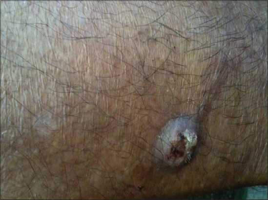
A 39-year- old male, farmer by occupation, presented with a single asymptomatic nodule with episodic discharge on right leg since six months which was gradually progressing in size (Fig. 1). There was no history of trauma and symptoms suggestive of systemic illnesses. On examination, there was a soft, non- tender mobile nodule of 1.5×1.5 cm with central ulceration on anteromedial aspect of right lower leg five cm above the medial malleolus. Initially, biopsy of the lesion showed mixed lymphohistiocytic infiltration with ill-defined granulomas in dermis and aggregates of neutrophils in epidermis. A provisional diagnosis of deep mycosis was made and the patient was started on Itraconazole. There was no response to treatment even after six months of itraconazole, and interestingly the patient developed two new lesions on flexors of left upper arm and right wrist and a satellite lesion adjacent to previously existing lesion. A repeat biopsy showed dense upper dermal infiltrate of lymphocytes and histiocytes with a narrow grenz zone (Fig. 2a) with positive Fite stain (Fig. 2b).Slit skin smear for bacteriological index from ear lobe came out to be 6+ positive. A final diagnosis of single nodular multibacillary leprosy without reaction and deformity was made. The patient was started on multidrug therapy as per WHO multi-bacillary regimen. There was gradual and complete resolution of lesions over a period of time.

Single nodulo-ulcerative lesion is a rare initial presentation of multibacillary leprosy. Classically lepromatous leprosy presents initially as macules, diffuse papules, infiltration,nodules or all four. Macules are small, multiple, erythematous or faintly hypopigmented, with shiny surface and vague border. Papules and nodules usually have normal skin color but with symmetrical distribution on face, arm, legs, and buttocks. In Polar LL, diffuse infiltrations may precede nodulation by many years. A part from these clinical presentations, lepromatous leprosy has been described in literatures with other forms of presentations which includes solitary plaques, urticarial wheals, adenoma sebaceum, and cheilitis granulomatosa [2–7].
Our case remained a diagnostic challenge initially because of low threshold of suspicion of leprosy because of pre -occupied notion of elimination of Leprosy, further confused by single nodular presentation of the disease. As the patient did not respond to antifungal therapy and developed new lesion on other site, a second biopsy was done which showed dermal infiltrate of lymphocytes and histiocytes with narrow grenz zone. This gave us the clue to diagnosis of leprosy, and was confirmed by positive slit skin smear, and demonstration of bacilli on Fite stain.
The advocacy of concerned authority for promoting health seeking behaviour for detection of leprosy in public in Nepal has so far been on identification of anaesthetic dry patch (i.e, more towards tuberculoid pole). We must have missed the lepromatous pole, which is more alarming, because of higher rates of transmission in this polar form.
More so, this case raises an important issue, whether we have been missing cases of lepromatous leprosy because of unusual presentation in post elimination era. To conclude, even though leprosy is on the verge of elimination, we as clinicians need to be vigilant about diagnosing a nodulo-ulcerative lesion and keep leprosy as one of the differential diagnoses of such presentations.
Consent
The examination of the patient was conducted according to the Declaration of Helsinki principles.
REFERENCES
1. Annual report. Department of health services. Government of Nepal, Ministry of health and population, Department of health services, Kathmandu, Nepal 2016/17: 149-162.p
2. Raut S, Kanade S, Nataraj G, Mehta P. Unusual presentation of multibacillary Leprosy. J lab Physic. 2017;9:57-9.
3. Armour KS, Scolyer RA, Barneston RS. Borderline Lepromatous Leprosy presenting as a single cutaneous plaque. Australas J Dermatol. 2005;46:181-83.
4. Sapkota BR, Neupane KD, Maharjan RK. Single lesion multibacillary leprosy, a treatment enigma: a case report. J Med Case Rep. 2009:3,8.
5. Kar BK, Belliappa PR, Ebenezer G, Job CK. Single lesion borderline lepromatous leprosy. Int J Lepros. 2004;72:45-7.
6. Booth AV, Kovich OI. Lepromatous leprosy. Dermatol Online J. 2007;13:9.
7. Barman KD, Gupta U, Saify K, Luthra S. Lepromatous leprosy with unusual features resembling adenoma sebaceum. Indian J Lepr. 2004;76:359-60.
Notes
Source of Support: Nil
Conflict of Interest: None declared.
Request permissions
If you wish to reuse any or all of this article please use the e-mail (brzezoo77@yahoo.com) to contact with publisher.
| Related Articles | Search Authors in |
|
|
|

Comments are closed.