|
|
Multiple seborrheic keratoses of the penis
Mohammed Chaouche , Selma El Kadiri, Younes Barbach, Abdellah Dah Cherif, Sara Elloudi, Hanane Baybay, Fatima Zahra Mernissi
Department of Dermatology and Venereology, University Hospital Hassan II, Fez, Morocco
Corresponding author: Dr. Mohammed Chaouche, E-mail: medch11@hotmail.com
Submission: 07.01.2019; Acceptance: 14.03.2019
DOI: 10.7241/ourd.2019e.13
Cite this article: Chaouche M, El Kadiri S, Barbach Y, Cherif AD, Elloudi S, Baybay H, Mernissi FZ. Multiple seborrheic keratoses of the penis. Our Dermatol Online. 2019;10(e):e13.1-e13.2.
Copyright information
© Our Dermatology Online 2019. No commercial re-use. See rights and permissions. Published by Our Dermatology Online.
Sir,
Seborrheic keratoses (SKs) are common, benign tumors which usually develop on the face, neck, and trunk as well as the extremities. SK can occur in the genital region which is a rare entity [1]. We report here a case of multiple SK lesions located on the genitals areas in a 34-year-old man.
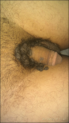
A 34-year-old man presented with multiple dark coloured growths over his genitals areas of 7 years duration. The lesion started as a small pigmented papule over the penis and left groin. Over the next few years, new asymptomatic lesions appeared over the adjacent areas. Past medical history was unremarkable and no history suggestive of risk of exposure to sexually transmitted diseases. Examination revealed multiple, black plaques, papules and nodules with lobulated, irregular, greasy surface (Fig. 1). Other mucocutaneous areas were normal and there was no lymphadenopathy. Systemic examination was non-contributory and human immunodeficiency virus tests were normal. Histopathological examination of biopsy specimens showed hyperkeratosis, papillomatosis and acanthosis with proliferation of basaloid cells containing multiple horn cysts. Dermis was unremarkable. Features were consistent with (SK). Shave excision of some lesions was done and the patient was reassured.
SKs are common benign tumor, which usually present with multiple pigmented papules and plaques. Lesions are rarely more than 1 cm in diameter and occur most often on trunk, face, and extremities, particularly over the sun exposed areas. Classically, SK tends to increase with the age in number and size. Morphologic variants of SK include the common flat type, skin tag like, stucco keratosis, dermatosis papulosa nigra, inverted follicular keratosis, and melanoacanthoma [1].
SK, can be easily differentiated by its characteristic histopathologic features. Several histological subtypes are generally recognized: Acanthotic, hyperkeratotic, adenoid, irritated, inverted follicular keratosis, and melanoacanthoma [2]. Of these, the acanthotic subtype is the most common variant. The acanthotic type, demonstrates hyperkeratosis, marked acanthosis with basaloid cells, papillomatosis and presence of horn cysts or pseudocysts [3]. The cause of genital SK is as yet unknown, but there may be a possible role of chronic friction [2]. Formation of in situ carcinoma and basal cell carcinoma has been documented rarely in acanthotic SK [2]. The common flat type of SK may be left alone or may be treated with liquid nitrogen cryotherapy, curettage, shave excision, or light electrodessication [4].
The genital area is rarely affected by seborrheic keratosis. This case report presented a rare form of seborrheic keratosis which affected the penis exclusively. This rare condition should be considered in the differential diagnosis for the lesions of the penis and histopathology after shave excision will help in the diagnosis.
Consent
The examination of the patient was conducted according to the Declaration of Helsinki principles.
REFERENCES
1. Quinn AG, Perkins W. Non-melanoma skin cancer and other epidermal skin tumours. In:Burns T, Breathnach S, Cox N, Griffiths C, editors. Rook's Textbook of Dermatology. 8th ed. Vol. 52. Oxford:Blackwell Science;2010. pp. 38–9.
2. Kirkham N. Tumors and cysts of the epidermis. In:Elder DE, Elenitsas R, Johnson BL, Murphy GF, editors. Lever's Histopathology of the Skin. 9th ed. Philadelphia:Lippincott Williams and Wilkins;2005. pp. 809–13.
3. El Amrani F, Meknassi I, Bouaddi M, Raffas W, Kettani F, Senouci K, et al. Giant seborrhoeic keratosis in an unusual site. Ann Dermatol Venereol. 2012;139:723–6.
4. Bandyopadhyay D, Saha A, Mishra V. Giant perigenital seborrheic keratosis. Indian Dermatol Online J. 2015;6:39–41.
Notes
Source of Support: Nil
Conflict of Interest: None declared.
Request permissions
If you wish to reuse any or all of this article please use the e-mail (brzezoo77@yahoo.com) to contact with publisher.
| Related Articles | Search Authors in |
|
 http://orcid.org/000-0003-3455-3810 http://orcid.org/000-0003-3455-3810
|


Comments are closed.