Giant melanoma on the scalp – clinical image
Piotr Brzeziński1,2, Józef Jasonek2, Mieczysława Karcz2, Joanna Kisiel2, Anna Możarowska2, Justyna Słomka2
1Institute of Biology and Environmental Protection, Pomeranian Academy, Slupsk, Poland, 2Department of Dermatology, Provincial Specialist Hospital in Slupsk, ul. Mickiewicza 12, 76-270 Ustka, Poland
Corresponding author: Dr. Piotr Brzezinski, E-mail: brzezoo77@yahoo.com
Submission: 20.12.2018; Acceptance: 02.01.2019
DOI: 10.7241/ourd.2019e.2
How to cite this article: Brzeziński P, Jasonek J, Karcz M, Kisiel J, Możarowska A, Słomka J. Giant melanoma on the scalp – clinical image. Our Dermatol Online. 2019;10(e):e2.1-e2.2.
Article in PDF file ![]()
The term “giant melanoma” is generally used to describe those cases of melanoma with a very large diameter independent of its depth [1,2]. There is no cutoff diameter for diagnosis of giant melanoma, although some authors confine this term to those lesions having a diameter larger than 10 cm [1,3].
The first description of a giant cutaneous melanoma dates back to 1970, when Bazex et al. described a case in a child [4].
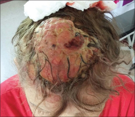
We present a case of giant melanoma of the scalp observed in our department by 63 years old women. It is a rare but very aggressive pathology which is generally treated by radical surgery. We emphasize early diagnosis because a large extension of the tumor can lead to the need of a very extensive surgical resection.
The lesion had appeared about few years before and it had progressively increased in size. At first, she neglected the problem, but sought medical attention after the mass began bleeding. Physical examinations revealed a malodorous, brown-reddish, firm, bleeding vegetative mass with necrotic areas and purulent exudation (Figs 1a and 1b). The dimension of the tumor was 11 cm x 10 cm x 4 cm. Mild erythema appeared in the surrounding skin. After confirming the melanoma in the histopathological examination, the patient was referred to the Oncological Surgery Department for palliative treatment.

Malignant melanomas extended from the scalp and neck have a particularly poor prognosis [5]. According to Grisham, a melanoma is defined as giant if its width is equal to or greater than 10 cm and its thickness reaches at least 4 mm [6].
Their growth is usually very rapid and their prognosis, if we associate them with other locations of melanomas classified as giants, is very reserved [7].
Panajotovic et al. [8] describe the case of a 57-year-old man with an exophytic pedicle 12X10 cm lesion corresponding to nodular melanoma ulcerated with a Breslow thickness of 100 mm. The patient does not have metastases or ganglia invaded despite tumor growth over three years.
His treatment was limited to surgical excision and the patient died of another pathology three weeks after the operation.
Ching and Gould [9] report the case of a 70-year-old man with a lesion that has been evolving for 3 months and measures 14.5×10.4 cm. It is exophytic, ulcerated corresponding to a grade 5 melanoma according to Clark and a Breslow thickness of 18mm.
The few cases described do not make it possible to release one or the other specificity concerning the very extensive melanomas of the scalp. It is therefore legitimate to associate them with other localizations of malignant melanomas of large size as to the therapeutic attitude.
The first treatment of these extensive lesions is the wide excision of the tumor with a margin of 2 to 3 cm.
Prognosis is very severe despite adjuvant medical treatments.
It is important to emphasize the importance of early diagnosis, which greatly facilitates surgery and other adjuvant treatments [10].
REFERENCES
1. di Meo N, Stinco G, Gatti A, Errichetti E, Bonin S, Albano A, et al. Giant melanoma of the abdomen: case report and revision of the published cases. Dermatol Online J. 2014;20.pii: 13030/qt4pp2825w.
2. Calteux N, Berchem G, Schmid N, Nebendahl J, Fischer G. [Giant melanoma of the scalp: Case report]. Bull Soc Sci Med Grand Duche Luxemb. 2013;(1):40-6.
3. Yuksel ME, Tamer F. Nodular malignant melanoma in the nasolabial fold. Our Dermatol Online. 2017;8:352-3.
4. Bazex A, Dupre A, Christol B, Cantala P, Carton. [Giant melanoma in children and a tumor with a sarcomatous structure]. Bulletin de la Societe francaise de dermatologie et de syphiligraphie. 1970;77:428-30.
5. Grisham AD Giant melanoma: novel problem, same approach. South Med J. 2010;103:1161-2.
6. Bebe FN, Hu S, Brown TL, Tulp OL. Metastatic melanoma in Florida, 1996-2010: Racial, demographic, occupational and tumor characteristics, and burden of metastasis. Our Dermatol Online. 2018;9:369-79.
7. Müller CS, Hinterberger L, Vogt T, Pföhler C. Giant melanoma of the scalp-discussion of a rare clinical presentation. BMJ Case Rep. 2011;2011.pii: bcr1220103643.
8. Panajotovic L, Dordevic B, Pavlovic MD. A giant primary cutaneous melanoma of the scalp – can it be that big? J Eur Acad Dermatol Venerol. 2007;21:1417-8.
9. Ching JA, Gould L. Giant Scalp Melanoma. A Case report and Review of the Literature. Eplasty. 2012;12:e51.
10. Smith N, Finn M, Segars L, Burns E, Peterson J, Sutton A, Vogt K, Menser M. Melanoma and medical education: knowledge and sun safety practices amongst medical students. Our Dermatol Online. 2018;9:11-4.
Notes
Source of Support: Nil
Conflict of Interest: None declared.
Comments are closed.