An angiomatous nodule of the cheek: What is your diagnosis?
Chaymae Jroundi , Sara Elloudi, Jihad Kassel, Zakia Douhi, Hanane Baybay, Fatima Zahra Mernissi
Department of Dermatology, University Hospital Hassan II of Fez, Morocco
Corresponding author: Chaymae Jroundi, MD
How to cite this article: Jroundi C, Elloudi S, Kassel J, Douhi Z, Baybay H, Mernissi FZ. An angiomatous nodule of the cheek: What is your diagnosis?. Our Dermatol Online. 2022;13(e):e40.
Submission: 02.02.2022; Acceptance: 01.06.2022
DOI: 10.7241/ourd.2022e.40
Citation tools:

Copyright information
© Our Dermatology Online 2022. No commercial re-use. See rights and permissions. Published by Our Dermatology Online.
CASE REPORT
A 35-year-old woman, with no particular history, presented with a lesion on the right cheek that appeared 8 years ago, progressively increasing in size over the past year, with the notion of bleeding to touch. The examination revealed a well-limited angiomatous nodule of 1.5 cm which was soft in consistency in a patient with skin phototype IV (Fig. 1). Dermoscopy: white structureless areas, polymorphic vascularization made of tortuous, linear and arborescent vessels, no pigment was observed (Figs. 2a and 2b). No cervical lymphadenopathy was found. A biopsy was performed showing: an ulcerated epidermis covering a dermis which is totally infiltrated by a proliferation made of nests of monomorphic medium sized cells, with eosinophilic cytoplasm, enlarged nuclei, a vesicular chromatin showing mitosis figures. A vascular reduced stroma was also found. An immunohistochemical study was necessary to determine the nature of the tumor showing a diffuse positive marking of the tumor cells with the following markers: Anti-HMB45 antibody, anti-PS100 antibody, anti-melan A antibody. A full-body CT scan didn’t find any abnormalities. The patient was scheduled for an excision of the tumor with appropriate margins.
 |
Figure 1: Well-limited angiomatous nodule of 1.5 cm on the right cheek. |
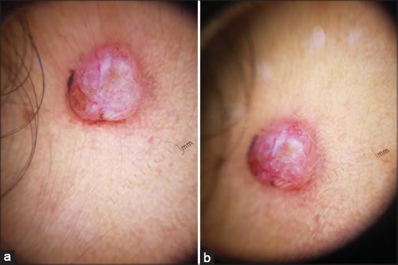 |
Figure 2: Dermoscopy: white structureless areas (a), polymorphic vascularization made of tortuous, linear and arborescent vessels, no pigment was observed (b). |
What’s your diagnosis?
Answer: amelanotic nodular melanoma
DISCUSSION
Herein we report a case of amelanotic nodular melanoma of the cheek. The absence of pigment and the chronicity of the lesion could lead to think of a pyogenic granuloma or a basal cell carcinoma. However, the recent change and the atypical appearance on dermoscopy led to suspect a more invasive and to perform a biopsy in the next day, which was in favor of a nodular melanoma. Amelanotic melanoma (AM) is a rare form of melanoma which lacks visible pigment [1]. AM represents 2% to 20% of all melanomas. It is thought to grow more rapidly than pigmented melanoma and to be associated with older age, freckling, and a sun-sensitive phenotype. (2) The difficulty of diagnosis lies in the lack of pigment, therefore clinical criteria like the ABCDE algorithm cannot be applied in this case [1,2]. The most common form is the nodular variant, but any other clinicopathological form can be found [3]. Clinical presentation varies from erythematous patch or plaque to angiomatous nodule or tumor in more advanced cases [1–3]. Although structures that suggest the presence of melanin are absent, dermoscopy has been a valuable tool in suspecting the diagnosis of AM [4]. A vascular pattern is the most reported with polymorphous vessels combined with milky-red globules or areas and/or red homogeneous areas [5]. The histopathological diagnosis of melanoma is usually evident. However, the histological appearance may simulate other lesions such as atypical fibroxanthoma or malignant fibrous histiocytoma, oriented by immunohistochemical study [6].
Although the cheek is a preferential site for the occurrence of achromic melanoma, our patient was still relatively young and did not have other cutaneous signs of sun damage. Dermoscopy has contributed to the orientation of the diagnosis and the adequate management in this case. Thus, an early dermatological evaluation and an appropriate dermoscopic examination may help avoid a delayed diagnosis. It is important to raise awareness among dermatologist to this rare and challenging entity.
ACKNOWLEDGEMENTS
The authors would like to acknowledge the patient.
Consent
The examination of the patient was conducted according to the principles of the Declaration of Helsinki.
The authors certify that they have obtained all appropriate patient consent forms, in which the patients gave their consent for images and other clinical information to be included in the journal. The patients understand that their names and initials will not be published and due effort will be made to conceal their identity, but that anonymity cannot be guaranteed.
REFERENCES
1. Kaizer-Salk KA, Herten RJ, Ragsdale BD, Sengelmann RD. Amelanotic melanoma:a unique case study and review of the literature. BMJ Case Rep. 2018;bcr-2017-222751.
2. Wee E, Wolfe R, Mclean C, Kelly JW, Pan Y. Clinically amelanotic or hypomelanotic melanoma:Anatomic distribution, risk factors, and survival. J Am Acad Dermatol. 2018;79:645-651.e4.
3. Silva TS, Araujo LR de, Faro GB de A, Paiva GR. Nodular amelanotic melanoma. An Bras Dermatol. 2019;94:497-8.
4. Lan J, Wen J, Cao S, Yin T, Jiang B, Lou Y, et al. The diagnostic accuracy of dermoscopy and reflectance confocal microscopy for amelanotic/hypomelanotic melanoma:a systematic review and meta-analysis. Br J Dermatol. 2020;183:210-9.
5. Pizzichetta MA, Kittler H, Stanganelli I, Ghigliotti G, Corradin MT, Rubegni P, et al. Dermoscopic diagnosis of amelanotic/hypomelanotic melanoma. Br J Dermatol. 2017;177:538-40.
6. Longvert C, Saiag P. Actualités dans le mélanome cutané. Rev Médecine Interne. 2019;40:178-83.
Notes
Source of Support: Nil,
Conflict of Interest: None declared.
Request permissions
If you wish to reuse any or all of this article please use the e-mail (brzezoo77@yahoo.com) to contact with publisher.
| Related Articles | Search Authors in |
|
|
 http://orcid.org/0000-0002-5942-441X http://orcid.org/0000-0003-3455-3810 http://orcid.org/0000-0002-5942-441X http://orcid.org/0000-0003-3455-3810 |

Comments are closed.