Eccrine poroma: When dermoscopy is useful for diagnosis
Madiha El Jazouly , Maha Salih Alj, FatimZahra Chahboun, Abderahman Al Bouzidi, Soumya Chiheb
1Dermatology Unit, Cheikh Khalifa International University Hospital, Mohammed VI University of Health Sciences, Casablanca, Morocco. 2Department of Anatomical and Cellular Pathology, Cheikh Khalifa International University Hospital, Mohammed VI University of Health Sciences, Casablanca, Morocco.
Citation tools:

Copyright information
© Our Dermatology Online 2022. No commercial re-use. See rights and permissions. Published by Our Dermatology Online.
ABSTRACT
Eccrine Poroma is a rare benign adnexal tumor. It presents the problem of differential diagnosis with several entities. Dermoscopic features are suggestive and may guide the diagnosis. We report a new observation. A 44-year-old woman, presented with an asymptomatic lesion on the dorsal side of the left foot for 2 years. Clinically the nodule was brownish, well delimited with superficial erosions, and firm on palpation. Dermoscopy showed linear and irregular vessels with perivascular white halos. Based on the clinical and dermoscopic features a diagnosis of eccrine poroma was made and confirmed by histological findings.
Key words: Eccrine poroma; Dermoscopy; Adnexal skin tumor
INTRODUCTION
Eccrine Poroma is a rare benign adnexal tumor. Usually, it is a solitary tumor, sessile or pedunculated papule or nodule from pink to red. Sometimes EP presents as a verrucous plaque and occasionally it can be pigmented. It appears more frequently in adults and commonly occurs on the soles and lateral aspects of the feet, but it can also be found on other anatomic sites such as the chest, eye, and buttocks [1]. Because of its clinical and dermoscopic variability, EP is usually difficult to recognize. It presents the problem of differential diagnosis with several entities. Dermoscopic features are suggestive and may guide the diagnosis. We report a new observation.
CASE REPORT
A 44-year-old woman, with no previous history of the disease, presented in our unit of dermatology with an asymptomatic lesion on the dorsal side of the left foot for 2 years. It gradually increased in size. Clinically the nodule was brownish, 0.8x1cm, well delimited with superficial erosions, and firm on palpation (Fig. 1). Clinical examination did not reveal similar lesions on the skin and the general examination including lymph nodes was normal. Differential diagnoses included squamous cell carcinoma, basal cell carcinoma, melanoma, epithelialized pyogenic granuloma, and histiocytofibroma. Dermoscopy showed linear and irregular vessels with perivascular white halos and whitish-pink areas with multiple purplish-red lacunae (Fig. 2). The nodule was completely excised and histopathologic examination showed a nodular proliferation of basaloid and squamoid cubic cells with vertical development in the epidermal extending to the deep dermis (Fig. 3). Based on the clinical, dermoscopic, and histological findings, a diagnosis of eccrine poroma (sensu stricto) was made.
 |
Figure 1: Well-circumscribed brownish nodule on the left foot. |
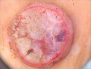 |
Figure 2: Dermoscopy: linear vessels (thin arrows), with perivascular white halos (thick arrows), red-blue lacunae (cercle), milky red areas (thick cross), whitich areas (triangle). (Polarized dermoscopy). |
 |
Figure 3: (a-b) Proliferation of basaloid and squamoid cubic cells with vertical development in the epidermal extending to the deep dermis (HE X 20). |
DISCUSSION
Eccrine poroma (Ep) was first described by Pinkus and al. in 1956. It is a rare benign adnexal tumor of epithelial cells originating from the terminal ductal portion of the sweat glands [2]. The pathogenesis of EP is largely unknown, but associations with scarring, radiation, and trauma have been reported. Clinically, it is usually a non-pigmented papular or nodular tumor located in the acral sites, which may be skin-colored, pink, red, or white. A pigmented variant (PEP) corresponds to 17% of cases [1,3]. It is usually confused with several potentially malignant lesions such as melanoma, squamous cell carcinoma, or pigmented basal cell carcinoma which gives it the character of the great clinical and dermoscopic imitator. More than one dermoscopic pattern has been described by many authors to characterize EP [4].
Therefore, dermoscopy becomes valuable and can significantly help distinguish an eccrine poroma from other tumors. In general, the most dermoscopic signs examined are the vascular structures to differentiate benign from malignant tumors. Dominguez. E and all concluded that the „chalice-form” and „cherry-blossom” vessels with a white-to-pink halo surrounding the vessels, and pink-white structureless areas are a good clue for the diagnosis of non-pigmented EP [5]. However, Ferrari and all observed that the vascular pattern is polymorphous including arborizing, hairpin, glomerular and linear vessels in both pigmented and non-pigmented poromas [6]. In our case, the pattern vessels are irregular and linear that clearly differed from the arborizing vessels usually seen in basal cell carcinoma. Other dermoscopic signs were observed, highlighting the absence of specific criteria for this benign tumor.
PEP dermoscopy is characterized by the presence of blue-gray ovoid nests, blue-gray dots, red-blue lacunae, and globule-like structures and other features such as the blue whitish veil and regression structures also reported. Thus, distinguishing the PEP from other malignant tumors such as basal cell carcinoma and melanoma or a benign tumor such as seborrheic keratosis remains difficult [7]. Minagawa and all suggested that the dermoscopic finding of reddish background colors which, histopathologically corresponds to the edematous stroma of poromas, may be an argument to differentiate poromas from melanocytic or other non-melanocytic pigmented tumors [8]. Bonbonato and all have used for examination a case of PEP, the dermoscopy, and the reflectance confocal microscopy (RCM). It has been useful to rule out the diagnosis of melanoma and the overall analysis of the architecture and cytology have suggested the diagnosis of an epithelial benign lesion [9].
CONCLUSION
In conclusion, although EP is characterized by multiple clinical and dermoscopic features and its definitive diagnosis is histopathological, dermoscopy and (RCM) remain promising tools to enhance the diagnosis, differentiate other tumors and avoid extensive surgery in some cases.
REFERENCES
1. Betti R, Bombonato C, Cerri A, Moneghini L, Abramo P, Menni S. Clinically and/or histologically pigmented poromas in Caucasian patients. G Ital Dermatol Venereol. 2014;149:341-6.
2. Goldman P, Pinkus, Rogin JR. Eccrine poroma;tumors exhibiting features of the epidermal sweat duct unit. AMA Arch Derm. 1956;74:511-21.
3. Elboukhari K, El Kadiri S, Benkirane S, Mernissi FZ. Adnexal benign tumor with deroupting dermoscopy. Our Dermatol Online. 2020;11:e87.1-e87.2.
4. Lallas A, Chellini PR, Guimares MG, Cordeiro N, Apalla Z, Longo C, et al. Eccrine poroma:the great dermoscopic imitator. J Eur Acad Dermatol Venereol. 2016;30:e61-e63.
5. Espinosa AE, Ortega BC, Venegas RQ, Ramírez RG. Dermoscopy of non-pigmented eccrine poromas:study of Mexican cases. Dermatol Pract Concept. 2013;3:25-8.
6. Ferrari A, Buccini P, Silipo V, De Simone P, Mariani G, Marenda S, et al. Eccrine poroma:a clinical-dermoscopic study of seven cases. Acta Derm Venereol. 2009;89:160-4.
7. Kuo HW, Ohara K. Pigmented eccrine poroma:a report of two cases and study with dermatoscopy. DermatolSurg. 2003;29:1076-9.
8. Minagawa A, Koga H. Dermoscopy of pigmented poromas. Dermatology. 2010;221:78-83.
9. Bombonato C, Piana S, Moscarella E, Lallas A, Argenziano G, Longo C. Pigmented eccrine poroma:dermoscopic and confocal features. Dermatol Pract Concept. 2016;6:59-62.
Notes
Request permissions
If you wish to reuse any or all of this article please use the e-mail (brzezoo77@yahoo.com) to contact with publisher.
| Related Articles | Search Authors in |
|
|
 http://orcid.org/0000-0003-2243-9615 http://orcid.org/0000-0003-2243-9615 |

Comments are closed.