Chronic intertrigo masking inverse psoriasis in an elderly female
Aida Oulehri , Zakia Douhi, Sara Elloudi, Hanane Baybay, Fatima Zahra Mernissi
Dermatology Department of the University Hospital Center Hassan II, Fez, Morocco
Corresponding author: Aida Oulehri, MD
How to cite this article: Oulehri A, Douhi Z, Elloudi S, Baybay H, Mernissi FZ. Chronic intertrigo masking inverse psoriasis in an elderly female. Our Dermatol Online. 2022;13(e):e18.
Submission: 11.09.2019; Acceptance: 11.12.2020
DOI: 10.7241/ourd.2022e.18
Citation tools:

Copyright information
© Our Dermatology Online 2022. No commercial re-use. See rights and permissions. Published by Our Dermatology Online.
ABSTRACT
Psoriasis is a chronic skin disorder that affects up to 5% of patients worldwide. The most common form of psoriasis is the chronic plaque type. Inverse psoriasis (IP) is characterized by its localization on inverse, intertriginous, or flexural body sites and is not always clinically obvious as the lesions usually lack the scale characteristic for typical psoriasis lesions, instead appearing smooth and shiny due to environmental factors. This clinical form is seen mostly in infants. In adults, it is much less frequent and has been suggested to be an indicator of an HIV infection. We report the case of a seventy-year-old female diagnosed with inverse psoriasis based on clinical and dermoscopic criteria. An assessment of the associations revealed no comorbidities.
Key words: Chronic intertrigo; Inverse psoriasis; Elderly
INTRODUCTION
Inverse psoriasis is a variety of psoriasis that involves the body folds, most often the axillary, anogenital, and inframammary. It is often difficult to diagnose due to its clinical similarity with other skin disorders involving the folds. Dermoscopy is a useful tool for enhanced non-invasive diagnosis. According to various studies and populations, the prevalence of IP is highly variable, ranging from 3% to 36% [1]. IP is typical in children, especially in young infants with involvement of the diaper area, configuring “napkin psoriasis” [2]. We report the case of a seventy-year-old female who had been, for three years, suffering from troublesome chronic intertrigo treated several times with oral and topical antimycotic agents but without improvement. We were able to diagnose reverse psoriasis with clinical methods and dermoscopy.
CASE REPORT
A seventy-year-old female with no notable pathological antecedents presented herself with pruritic lesions of the submammary and inguinal folds, which had been present for three years, for which she had consulted several times, and which were treated locally and topically as mycotic intertrigo without improvement. The lesions had evolved with flare-ups.
A clinical examination revealed well-limited, fine, and scaly erythematous plaques located in the submammary, subabdominal, and inguinal folds with extension to the labia majora (Figs. 1 and 2). Dermoscopy revealed scales with homogeneously distributed dotted vessels on a light-red background (Fig. 3). The rest of the examination found scaly patches on the scalp with positive Auspitz’s sign on dermoscopy (Figs. 4a and 4b). In light of these findings, we maintained the diagnosis of IP and treated the patient with mid-potency topical steroids with great improvement. We also performed a metabolic workup as well as an HIV serology. All test results were negative.
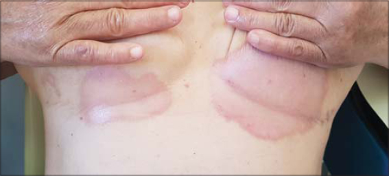 |
Figure 1: Well-limited, fine, and scaly erythematous plaques located in the submammary folds. |
 |
Figure 2: Well-limited, fine, and scaly erythematous plaques located in the subabdominal and inguinal folds with extension to the labia majora. |
 |
Figure 3: Dermoscopy of the inguinal fold showing scales with homogeneously distributed dotted vessels on a light-red background. |
 |
Figure 4: (a) Scaly patches on the scalp with (b) positive Auspitz’s sign on dermoscopy. |
DISCUSSION
Localization of psoriatic lesions in the intertriginous areas affects the clinical presentation. The most evident difference between classic, plaque-type psoriasis and inverse psoriasis is the lack of, or less, scaling in the intertriginous areas. The lesions are well-demarcated, erythematous, and often presenting with a shiny appearance [1]. IP is difficult to diagnose due to its clinical similarity with other skin disorders involving the folds, mainly mechanical intertrigo, fungal and bacterial infections, contact dermatitis, seborrheic dermatitis, and lichen planus [3]. The diagnosis of PI is most often clinical, and a complete examination of the entire tegument, including the scalp, nails and mucous membranes, is important and may offer arguments in favor of psoriasis. This was the case in our patient, in whom scaly patches of the scalp, which were not mentioned during questioning, were found. Dermoscopy of plaque psoriasis typically shows a characteristic pattern consisting of diffuse white scales and symmetrically and regularly distributed dotted vessels on a light or dull red background [4]. It is a very useful tool for this topographic form and was a great help to us in rectifying the diagnosis of our patient.
Psoriasis, in general, has been associated with comorbidities such as cardiovascular disease, metabolic syndrome, and lymphoma [1]. Currently, no work has been published on comorbidities associated with inverse psoriasis. The development of inverse psoriasis has been reported as a paradoxical side effect to treatment with infliximab for Crohn’s disease [5] and hidradenitis suppurativa [6]. Inverse psoriasis might be more common in people with HIV, possibly indicating a different immunologic interplay [7]. A tendency toward overrepresentation of inverse psoriasis in the obese could suggest friction or maceration as a triggering factor of psoriasis in intertriginous sites as well [8]. From a pharmacological point of view, application of topical treatments in the intertriginous areas may be considered as a treatment under occlusion due to enhanced hydration and increased percutaneous absorption. The inverse areas are considered more sensitive and prone to side effects from topical steroids. Treatment with steroids should be prescribed with caution to avoid side effects [1]. The current short-term first-line treatment for inverse psoriasis is low- or mid-potency topical steroids [9].
CONCLUSION
Inverse psoriasis is considered an infrequent site of involvement of psoriasis. It is sometimes difficult to diagnose due to its clinical similarity with other skin disorders involving the folds. Dermoscopy is a useful tool for enhanced non-invasive diagnosis. Its treatment may be challenging as intertriginous areas are more prone to side effects induced by topical therapies. Several co-morbidities may be found, but may also be absent, as our case shows.
Consent
The examination of the patient was conducted according to the principles of the Declaration of Helsinki.
The authors certify that they have obtained all appropriate patient consent forms, in which the patients gave their consent for images and other clinical information to be included in the journal. The patients understand that their names and initials will not be published and due effort will be made to conceal their identity, but that anonymity cannot be guaranteed.
REFERENCES
1. Omland SH, Gniadecki R. Psoriasis inversa:A separate identity or a variant of psoriasis vulgaris?Clin Dermatol. 2015;33:456-61.
2. Bronckers IMGJ, Paller AS, van Geel MJ, van de Kerkhof PCM, Seyger MMB. Psoriasis in children and adolescents:Diagnosis, management and comorbidities. Paediatr Drugs. 2015;17:373-84.
3. Micali G, VerzìAE, Giuffrida G, Panebianco E, Musumeci ML, Lacarrubba F. Inverse psoriasis:From diagnosis to current treatment options. Clin Cosmet Investig Dermatol. 2020;12:953-9.
4. Errichetti E, Stinco G. Dermoscopy in general dermatology:A practical overview. Dermatol Ther (Heidelb). 2016;6:471-507.
5. Peramiquel L, Puig L, Dalmau J, Ricart E, Roe E, Alomar A. Onset of flexural psoriasis during infliximab treatment for Crohn’s disease. Clin Exp Dermatol. 2005;30:713-4.
6. Nuño-González A, Dehesa L, Ricotti C, Kerdel F. Flexural or inverse psoriasis in a patient with hidradenitis suppurativa receiving treatment with infliximab. Actas Dermosifiliogr. 2012;103:936-7.
7. Castillo RL, Racaza GZ, Roa FDC. Ostraceous and inverse psoriasis with psoriatic arthritis as the presenting features of advanced HIV infection. Singapore Med J. 2014;55:e60-3.
8. Herron MD, Hinckley M, Hoffman MS, Papenfuss J, Hansen CB, Callis KP, et al. Impact of obesity and smoking on psoriasis presentation and management. Arch Dermatol. 2005;141:1527-34.
9. Reynolds KA, Pithadia DJ, Lee EB, Wu JJ. Treatments for inverse psoriasis:A systematic review. J Dermatolog Treat. 2020;31:786-93.
Notes
Source of Support: Nil,
Conflict of Interest: None declared.
Request permissions
If you wish to reuse any or all of this article please use the e-mail (brzezoo77@yahoo.com) to contact with publisher.
| Related Articles | Search Authors in |
|
|
 http://orcid.org/0000-0002-5942-441X http://orcid.org/0000-0003-3455-3810 http://orcid.org/0000-0002-5942-441X http://orcid.org/0000-0003-3455-3810 |

Comments are closed.