Heterochromia of beard hair – A rare case report
Vijeta Prasad, Alpana Mohta , Rekha Srinivasa, Bhikamchand Ghiya
Department of Dermatology, Venereology and Leprology, Sardar Patel Medical College, Bikaner, Rajasthan, India
Corresponding author: Alpana Mohta, MD
How to cite this article: Prasad V, Mohta A, Srinivasa R, Ghiya B. Heterochromia of beard hair – A rare case report. Our Dermatol Online. 2022;13(e):e14.
Submission: 28.09.2021; Acceptance: 17.01.2022
DOI: 10.7241/ourd.2022e.14
Citation tools:

Copyright information
© Our Dermatology Online 2022. No commercial re-use. See rights and permissions. Published by Our Dermatology Online.
ABSTRACT
Heterochromia of hair is defined as presence of different colour of hair in a same individual. The colour of hair can vary from red,brown, yellow to white.The distribution of heterochromic hair could be symmetric and asymmetric. Distribution pattern can also be described as patchy,diffuse and segmental. The segmented heterochromia of scalp hair is characterized by alternate light and dark segment which is seen in iron deficiency anemia. Diffuse scalp heterochromia that is even distribution on whole scalp may be associated with systemic disease like phelyketonuria or kwashiorkor, certain medications and could be simply physiological or familial. It is very rare identity and there has been very few case reports on heterchromia of scalp hair. Here, we report a unique case of 24 year old male who presented with heterochromia of beard hair.
Key words: Heterochromia; Beard; hair
INTRODUCTION
Heterochromia of hair is a rare finding. The distribution of heterochromic hair can be patchy, diffuse, segmental and blashkoid [1,2]. It could be present at birth or develop later in life. The case we are presenting here has heterochromia of beard which was not present initially but acquired later in life.
CASE REPORT
A 24 year old male patient presented to our skin out-patient department with red-brown discolouration of beard hair present diffusely within black hair symmetrically on both sides of face (Figs. 1a and 1b). The hair over scalp, eyebrows, eyelashes, axillary, pubic and body hair were black in colour. General physical examination was normal. Cutaneous and mucosal examination was normal. No abnormalities were detected in teeth and nails. On inquiry, patient revealed that the hair were initially black in color and had turned into a lighter color only within the last 1 year. There was no history of any topical drug application, systemic drug use, any hair dye, bleaching agent or chemical exposure, trauma or inflammatory disease prior to onset of lesions. The routine blood investigations-CBC, RFT, LFT, urine microscopy and serum ferritin, vitamin B12, serum calcium, vitamin D, thyroid profile, serum zinc, serum copper levels were within normal limits. There was no similar history of heterochromic hairs in family and birth out of consanguinity. On dermatoscopic examination the brownish red hair were similar in width and length to black hair (Fig. 2). The pigmentation was uniform across the entire length of hair. The case was clinically diagnosed as heterochromia of beard hair. Molecular genetic analysis for pigmentary mosaicism was not done.
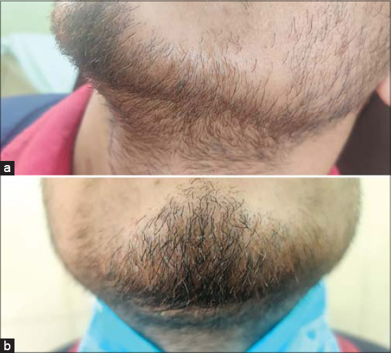 |
Figure 1: a and b – Clinical photograph showing symmetrical heterochromia of beard (a side view & b frontal view). |
 |
Figure 2: Dermatoscopic image showing uniform reddish brown pigmentation of hair distributed in between black hair (Dermlite DL4 & 3 Gen10x). |
The counselling of patient regarding benign nature of this condition was done and no treatment was given. The patient was followed up regularly for next 1 year. During this period there was no progression of the condition and no further change in colour of already existing reddish brown hair found.
DISCUSSION
The process of follicular melanogenesis is connected with follicular cycling and melanin synthesis occurs only during anagen phase. The type of melanosome present in hair follicle melanocytes determines the colour of hair. The black hair follicle has highest number of eumelanosomes, whereas, red hair follicle has pheomelanosomes. The genetic control of hair colour is thought to be polygenic in inheritance. Heterochromia can be present in symmetrical and asymmetrical pattern. Asymmetrical pattern signifies genetic basis [2,3]. Distribution pattern can also be described as patchy, diffuse and segmental. The segmented heterochromia of scalp hair is characterized by alternate light and dark segment which is seen in iron deficiency anemia. Diffuse scalp heterochromia that is even distribution on whole scalp may be associated with systemic disease like phelyketonuria or kwashiorkor, certain medications like dizoxide causing red hair, minoxidil causing black hair or chloroquine causing white hair and it could be simply physiological or familial without any underlying cause. There are various etiologies described in literature responsible for heterochromia like poliosis, white forelock of piebaldism, hairy melanocyctic nevus, vitiligo, tuberous-sclerosis, Waardenberg syndrome, metabolic disorders, drugs, autoimmune disease and nutritional deficiencies [4]. Halo nevus- rare dermatological entity of halo around melanocytic nevus is also reported to occur with poliosis [5,6]. Parry–romberg syndrome is characterized by atrophy, slowly progressive in nature involving the skin, fat and connective tissue located on one side of the face, affected individuals can also develop abnormalities like whitening of hair [7].
There are case reports of heterochromia of scalp hairs but to our best knowledge till now there has been no case reported so far of isolated heterochromia of beard hair. Under reporting of such presentation might be due to asymptomatic nature of this condition with no pigmentary mosaicism of skin causing no cosmetic concern. Thus, we are reporting this case due to rarity of its occurrence. However, a dermatologist must always be on the lookout to differentiate it from other similar spotters.
Consent
The examination of the patient was conducted according to the principles of the Declaration of Helsinki.
The authors certify that they have obtained all appropriate patient consent forms, in which the patients gave their consent for images and other clinical information to be included in the journal. The patients understand that their names and initials will not be published and due effort will be made to conceal their identity, but that anonymity cannot be guaranteed.
REFERENCES
1. Yoon KH, Kim D, Sohn S, Lee WS. Segmented heterochromia in scalp hair. JAm Acad Dermatol. 2003;49:1148–50.
2. Iorizzo M, Piraccini BM, Tosti A. Heterochromia of the scalp hair following Blaschko lines. Pediatr Dermatol. 2007;24:69–70.
3. Qiao J, Fang H. Heterochromia of the scalp hair. CMAJ. 2010;182:E862.
4. Dumitrascu CI, Hoss E, Hogeling M. Heterochromia of the hair and eyelashes with blaschkoid dyspigmentation. Pediatr Dermatol. 2016;33:e121-2.
5. Achehboune K, Zinoune S, Baybay H, Elloudi S, Mernissi FZ. Halo nevus in the dermoscopy. Our Dermatol Online. 2020;11:e46.1-2.
6. Fellman AC, Mehregan AH. Halo Nevi of Scalp With Poliosis. Arch Dermatol. 1976;112:559.
7. Ramalingam A, Sreedevi A. A rare case of Parry Romberg syndrome with morphea. Our Dermatol Online. 2020;11:e133.1-3.
Notes
Source of Support: Nil,
Conflict of Interest: None declared.
Request permissions
If you wish to reuse any or all of this article please use the e-mail (brzezoo77@yahoo.com) to contact with publisher.
| Related Articles | Search Authors in |
|
|
 http://orcid.org/0000-0001-7526-2089 http://orcid.org/0000-0001-7526-2089 |

Comments are closed.