Verrucous hemangioma – Rare case report
Muddennavar Snehal , Selvakumar Monishadevi, Patra Anjankumar
Department of Dermatology, MVJ Medical College and Research Hospital, Bangalore, India
Corresponding author: Muddennavar Snehal, MD
How to cite this article: Snehal M, Monishadevi S, Anjankumar P. Verrucous hemangioma – Rare case report. Our Dermatol Online. 2022;13(e):e12.
Submission: 09.06.2021; Acceptance: 23.01.2022
DOI: 10.7241/ourd.2022e.12
Citation tools:

Copyright information
© Our Dermatology Online 2022. No commercial re-use. See rights and permissions. Published by Our Dermatology Online.
ABSTRACT
Verrucous hemangioma is an uncommon localized,congenital vascular malformation of the cutaneous and subcutaneous tissues with a predilection for the lower extremities. Here we present a varied clinical presentation.44 year old female presented with dark raised itchy lesion over the right knee since birth, non progressive associated with itching more in the night. O/E: Multiple,well-defined, hyperpigmented, hyperkeratotic plaques with minimal scaling over right knee and right thigh. Doppler study showed multiple ectatic varicosities and skin biopsy showed ectatic blood vessels. Verrucous hemangioma typically starts as a bluish red lesion with small satellites. Lesions are characteristic by bluish black color and develop a verrucous surface. MRI is the diagnostic modality of choice because it delineates the planes and guides biopsy. Histopathological examination remains the gold standard for diagnosis. Verrucous hemangiomas do not resolve spontaneously and early diagnosis is important for timely surgical excision and better cosmetic result.
Key words: Verrucous hemangioma; Cavernous hemangioma; Angiokeratoma; Lymphangioma circumscriptum
INTRODUCTION
Verrucous hemangioma is a rare, congenital, superficial angiokeratoma-like vascular malformation composed of capillaries and veins in the dermis and upper subcutis with a predilection for the lower extremities and spreads slowly [1]. As it spreads, disseminated nodules may develop [2]. Differential diagnosis includes angiokeratoma, lymphangioma circumscriptum, verrucous epidermal nevus, or even malignant melanoma [3]. The condition is observed at birth or in early childhood. Initially it presents as bluish-red in color. Later the lesions acquire a verrucous or warty surface. Secondary infection, a frequent complication, results in reactive papillomatosis and hyperkeratosis. Unlike other angiomatous nevi, it does not involute spontaneously. Verrucous haemangioma was first described in 1967 by Imperial and Hedwig. Here we present a varied clinical presentation of Verrucous Hemangioma [4]. We describe a rare case of verrucous hemangioma in a middle aged woman with numerous warty lesions over the right knee since birth.
CASE REPORT
The patient was a 44 year old female presented with dark raised warty lesion over the right knee since birth, non progressive associated with itching more in the night.
History of bleeding usually following minor trauma was noted.
No history of trauma/fever
General Physical Examination: No abnormality detected.
Cutaneous examination:
Multiple, well defined, hyperpigmented, hyperkeratotic plaques with overlying minimal scaling, largest measuring 7x5cms over the right knee and smallest measuring 2x2cm over the right thigh. Surface of the lesion was firm and verrucous with no ulceration, bleeding, or atrophy. The lesion was non compressible and diascopy was negative (Figs. 1a–1c).
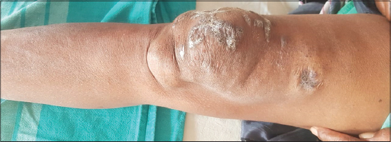 |
Figure 1a: Multiple, well defined, hyperpigmented , hyperkeratotic plaques with overlying minimal scaling. Largest lesion measuring. |
 |
Figure 1b: 7x5cms over the right knee and smallest measuring 2x2cm over the right thigh. Surface of the lesion was firm and verrucous surface. |
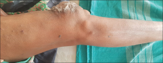 |
Figure 1c: Solitary well defined swelling, soft in consistency, non tender, pinchable present below right knee. |
Solitary well defined swelling of size 5x7cm,soft in consistency, non tender, pinchable present below right knee. Hair/Nail/Mucosa – Normal. There was no palpable regional lymphadenopathy.
Doppler study showed multiple ectatic varicosities in right knee and skin biopsy showed ectatic blood vessels extending from the papillary dermis into the subcutaneous tissue, diagnostic of verrucous hemangioma. MRI could not be done due financial constraints. Hence patient was treated symptomatically and was referred to vasucular surgeon for further management but patient lost to follow up.
DISCUSSION
Verrucous hemangioma is an uncommon, localized, congenital vascular malformation [4]. It initially appears as a bluish macule that later gets an erythematous-violaceous colour, and following trauma and secondary infections, often evolves into a verrucous plaque/nodule [5]. Clinically, lesions may simulate angiokeratoma, lymphangioma circumscriptum, verrucous epidermal nevus, or even malignant melanoma. MRI is the diagnostic modality of choice because it delineates the dermal and subcutaneous plane of these lesions and guides biopsy. Histopathological examination remains the gold standard for diagnosis [6]. Early diagnosis is important to obtain an excellent cosmetic outcome. Verrucous hemangioma should be excised while still small to prevent scars. The treatment of choice for verrucous hemangiomas is wide and deep surgical excision in order to ensure free margins to reduce the chance of possible recurrences, as a superficial therapeutic approach would be unsuccessful because of the deep angiomatous proliferation. Cryosurgery, electrocautery, or laser therapy can be used [7].
CONCLUSION
To conclude, we report this case for its rarity of occurrence and its clinical implications for the differential diagnosis of various conditions, including melanoma, as described above. Verrucous hemangiomas do not resolve spontaneously and early diagnosis is important for timely surgical excision and better cosmetic result.
Consent
The examination of the patient was conducted according to the principles of the Declaration of Helsinki.
The authors certify that they have obtained all appropriate patient consent forms, in which the patients gave their consent for images and other clinical information to be included in the journal. The patients understand that their names and initials will not be published and due effort will be made to conceal their identity, but that anonymity cannot be guaranteed.
REFERENCES
1. Boccara O, Ariche-Maman S, Hadj-Rabia S, Chrétien-Marquet B, Frassati-Biaggi A, Zazurca F, et al. Verrucous hemangioma (also known as verrucous venous malformation):A vascular anomaly frequently misdiagnosed as a lymphatic malformation. Ped Dermatol. 2018;35:e378-81.
2. Bindhuja J, Rajendiran S, Priyathersini N, Balaji Singh K, Leena DJ. Verrucous hemangioma:a rare vascular tumor –a case report. Sri Ramachandra J Med. 2013;6:2021.
3. Chung WJ, Chung HW, Shin MJ, Lee SH, Lee MH, Lee JS, et al. MRI to differentiate benign from malignant soft-tissue tumours of the extremities:a simplified systematic imaging approach using depth, size and heterogeneity of signal intensity. Br J Radiol. 2012;85:e831-6.
4. Singh J, Sharma P, Tandon S, Sinha S. Multiple verrucous hemangiomas:A case report with new therapeutic insight. Indian Dermatol Online J. 2017;8:254.
5. Mestre T, Amaro C, Freitas I. Verrucous haemangioma:a diagnosis to consider. Case reports. 2014;2014:bcr2014204612.
6. Nagarajan K, Banushree CS. Usefulness of MRI in delineation of dermal and subcutaneous verrucous hemangioma. Indian J Dermatol. 2015;60:525.
7. Fatani M, Al Otaibi H, Mohammed M, Hegazy O. Verrucous hemangioma treated with electrocautery. Case Rep Dermatol. 2016;8:112-7.
Notes
Source of Support: Nil,
Conflict of Interest: None declared.
Request permissions
If you wish to reuse any or all of this article please use the e-mail (brzezoo77@yahoo.com) to contact with publisher.
| Related Articles | Search Authors in |
|
|
|

Comments are closed.