Cutaneous leishmaniasis: A rare possibility of discovering HIV
Hind Palamino , Zoubida Mehssass, Fatima Azzahrae El Ghaitibi, Mariame Meziane, Nadia Ismaili, Leila Benzekri, Karima Senouci
Dermatology-Venereology Department,CHU Ibn sina, Rabat, Morroco
Corresponding author: Palamino Hind, MD
Submission: 06.11.2020; Acceptance: 23.01.2021
DOI: 10.7241/ourd.2021e.53
Cite this article: Palamino H, Mehssass Z, El Ghaitibi FA, Meziane M, Ismaili N, Benzekri, L, Senouci K. Cutaneous leishmaniasis: A rare possibility of discovering HIV. Our Dermatol Online. 2021;12(e):e53.
Citation tools:
Copyright information
© Our Dermatology Online 2021. No commercial re-use. See rights and permissions. Published by Our Dermatology Online.
Sir,
Leishmaniasis is a disease caused by the flagellated protozoa Leishmania spp. It is frequently responsible for cutaneous and visceral lesions. Several cases of cutaneous and visceral leishmaniasis in HIV-positive patients have been described, but cases of cutaneous leishmaniasis (LC) revealing HIV have been rarely reported [1,2].
We report two cases of young patients, in otherwise good health, with chronic ulcerations revealing HIV coinfections and cutaneous leishmaniasis.
Case 1: A 28-year-old male from Ouazzane, an endemic area in Morocco, consulted for a spontaneously painful chronic ulceration at the internal malleolus, which had been evolving for around one year.
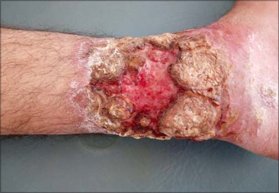
A clinical examination revealed an ulceration on the left internal malleolus: bilateral, mobile, smelly, crusty, and painful with a serofibrinous background and inflammatory edges, 10 cm in diameter by 8 cm (Fig. 1), with inguinal lymphadenopathy and a firm infracentimetric epitrochlea.
 |
Figure 1: Ulceration of the medial malleolus with a serofi brinous background and a crusty border surrounded by an infl ammatory halo. |
The patient did no hepatomegaly, splenomegaly, or signs of chronic venous insufficiency. A biological assessment was unremarkable. An arterial and venous Doppler ultrasound of the lower extremities and radiography of the leg were unremarkable. A smear from the lesion revealed the presence of Leishman’s bodies under microscopy (Fig. 2).
 |
Figure 2: Microscopy revealing amastigotes. |
A skin biopsy revealed lymphohistiocytic infiltration, suggesting leishmaniasis. An abdominopelvic ultrasound was normal. The patient refused a sternal puncture.
Faced with the chronic and extensive nature of the lesion and the presence of lymphadenopathy, we completed our assessment with an HIV serology, which was positive. The CD4 T cell count was 112/mm³.
Due to lack of resources, we were unable to determine the species of the leishmania.
The patient was treated with the pentavalent antimonial meglumine antimoniate (Glucantime®) 20 mg/kg/day intramuscularly for 21 days and triple therapy (Efavirenz, Emtricitabine, Tenofovir) combined with local care and prophylactic antibiotic therapy for opportunistic infections. The evolution was favorable.
Case 2: A 39-year-old male from Errachidia with no medical history and otherwise in good health consulted for ulcerated lesions on both forearms evolving for four months.
A clinical examination revealed two circumferential ulcerated lesions at both wrists: painless, well-limited, and with flexible erythematous edges. In the center of the right lesion, there was an ulcerated erythematous nodule 3 cm in diameter.
The rest of the clinical examination as well as a biological assessment were normal (Fig. 3).
 |
Figure 3: Two well-limited circumferential ulcers on both wrists with an ulcerated erythematous nodule on the right wrist. |
A smear of the lesion revealed the presence of Leishman’s bodies under microscopy (Fig. 2).
An ultrasound of the lymph nodes and thoracic, abdominal, and pelvic CT scans were normal and revealed no visceral localization.
In view of the extensive and chronic nature of the lesions, retroviral serology was performed. The patient was HIV-positive and the CD4 count revealed a stage IV disease (with the CD4 count at 89/mm3).
We initiated treatment on a pentavalent antimonial (meglumine antimonate) at a dose of 20 mg/kg/day for 21 days intramuscularly combined with the triple therapy.
The outcome was favorable and complete healing was achieved within two months (Fig. 4). We report two cases of atypical cutaneous leishmaniasis without visceral localization revealing an HIV infection in otherwise perfectly healthy, asymptomatic patients.
 |
Figure 4: Complete healing of the lesions after treatment. |
Leishmaniases are a group of infectious diseases that are widespread throughout the world. They are caused by the flagellated protozoa Leishmania spp. It is transmitted by the bite of an infected female sandfly of the genus Phlebotomus in the old world and the genus Lutzomyia in the new world [1,2].
In HIV-positive patients, the risk of contracting leishmaniasis is increased by 100 to 1000 times [3] and may contribute to the development of atypical clinical forms, with visceralization of dermotropic species, or to the cutaneous dissemination of species with visceral tropism [4].
Both leishmania and HIV multiply in monocytes. Myeloid cells infected with leishmaniasis facilitate HIV replication, while HIV promotes the phagocytosis of the leishmania by macrophages and amplifies parasitic replication in monocytes, which explains the clinical polymorphism and the association with general signs [5].
In our two cases, cutaneous leishmaniasis manifested itself as isolated chronic skin ulcerations without clinical signs suggesting an HIV infection. This illustrates the clinical polymorphism in such coinfection and the advantage of proposing HIV serology in atypical presentations of cutaneous leishmaniasis.
The recommended medicine is pentavalent antimonial derivatives at 20 mg/kg/day intramuscularly or intravenously for 10 to 20 days alone or in combination with pentoxifylline 400 mg 3 times a day for 10 to 20 days or fluconazole 200 mg/day orally for 6 weeks. In the absence of a response, intravenous liposomal amphotericin B at 1–1.5 mg/kg/day for 21 days or 3 mg/kg/day for 10 days may be proposed as a second-line treatment [1–6].
We chose a pentavalent antimonial alone for our two patients and observed a good evolution.
It must be noted that a low count of CD4 T lymphocytes (less than 200/mm3) exposed the patients to a risk of relapse and the onset of immune restoration syndrome [4]. However, although our patients had a low CD4 count, no relapses were noted after three years and two years, respectively.
Our observations reveal the benefit of performing HIV serology in patients with atypical cutaneous leishmaniasis, even in the absence of a secondary location or general signs.
Statement of Human and Animal Rights
All the procedures followed were in accordance with the ethical standards of the responsible committee on human experimentation (institutional and national) and with the 2008 revision of the Declaration of Helsinki of 1975.
Statement of Informed Consent
Informed consent for participation in this study was obtained from all patients.
REFERENCES
1. World Health Organization. Control of the leishmaniases. Geneva:World Health Organization. http://whqlibdoc.who.int/ trs/WHO_TRS_949_eng.pdf
2. Paudel V, Parajuli S, Chudal D. Cutaneous leishmaniasis in Nepal:An emerging public health concern. Our Dermatol Online. 2020;11:76-8.
3. Vasudevan B, Bahal A. Leishmaniasis of the lip diagnosed by lymph node aspiration and treated with a combination of oral ketaconazole and intralesional sodium stibogluconate. Indian J Dermatol. 2011;56:214-6.
4. Diatta BA, Diallo M, Diadie S, Faye B, Ndiaye M, Hakim H, et al. Cutaneous leishmaniasis due to Leishmania infantum associated with HIV:A case report. Ann Dermatol Venereol. 2016;143:625-8.
5. Talat H, Attarwala S, Saleem M. Cutaneous leishmaniasis with HIV. J Coll Physicians Surg Pak. 2014;24 Suppl 2:S93-5.
6. Buffet PA, Rosenthal É, Gangneux JP, Lightburne E, Coup-piéP, Morizot G, et al. Therapy of leishmaniasis in France:Consensuson proposed guidelines. Presse Med. 2011;40:173-84.
Notes
Source of Support: Nil,
Conflict of Interest: None declared.
Request permissions
If you wish to reuse any or all of this article please use the e-mail (brzezoo77@yahoo.com) to contact with publisher.
| Related Articles | Search Authors in |
|
|


Comments are closed.