Breast ulcerations: The dermatologist’s point of view through two observations
Meryem Khalidi , Achraf Machan, Mohammed El Amraoui, Naoufal Hjira, Mohammed Boui
Dermatology-Venerology Department at Mohammed V Military Hospital of Instruction, Rabat, Morrocco
Corresponding author: Meryem Khalidi, MD
Submission: 02.01.2021; Acceptance: 19.03.2021
DOI: 10.7241/ourd.2021e.49
Cite this article: Khalidi M, Machan A, El Amraoui M, Hjira N, Boui M. Breast ulcerations: The dermatologist’s point of view through two observations. Our Dermatol Online. 2021;12(e):e49.
Citation tools:

Copyright information
© Our Dermatology Online 2021. No commercial re-use. See rights and permissions. Published by Our Dermatology Online.
ABSTRACT
Breast ulcerations are a source of concern for dermatologists and also gynecologists. They can reveal unexpected various pathologies. We shed light on this entity through two observations of often unrecognized mammary dermatosis. The first case is about 24 years old female patient who consulted for ulcerative lesions of the breast and in whom the skin biopsy found an epitheloid and gigantocellular granuloma without caseous necrosis. The paraclinical examinations were able to retain the diagnosis of idiopathic granulomatous mastitis. The second case is about a 38 years old patient who presented a multiple non specific and erosive plaques breast which led to the diagnosis of pathomimia. These diagnoses were retained after eliminating several differential diagnoses and having recourse to therapeutic tests as well. Breast ulcerations are often sources of diagnostic difficulties, the reflex being to always think of an underlying mammary pathology, but certain purely dermatological conditions can advance the diagnosis
Key words: Breast ulcerations; Idiopathic granulomatous mastitis; Pathomimia
INTRODUCTION
The breast is made up of skin and connective tissue, encompassing the mammary gland. The latter derives from the sweat glands, given its functional role in lactation, its character as a sexual organ, and the frequency of breast cancer. Any breast disease is a source of significant anxiety for the patient. Between affections of the mammary gland fistulized to the skin, and ulcerated skin conditions threatening the mammary gland, both the dermatologist and the gynecologist are in difficulty. We report two observations of breast ulcerations where dermatological analysis was of great help in labeling the diagnosis.
CASE REPORTS
Case 1
24-year-old patient, who gave birth 1 year ago, and presented with non-regressive right breast engorgement, which prevented breastfeeding through that breast. She was seen in consultation for ulcerative budding, infiltrated, erythematous-purplish, suppurative and painless lesions of the right breast, progressing for 2 months, with inflammatory arthralgia and erythema nodosum (Fig. 1). Biologically, the patient had presented a moderate inflammatory syndrome. Pus swabs with bacteriological examination for typical and atypical germs, as well as mycological examinations, were negative. The search for the koch bacillus genome by polymerase chain reaction on pus and tissue samples remained negative. An MRI of the breast revealed the presence of multiple breast collections from the right breast, fusing towards the axillary extension and towards the infero-external quadrant, the largest measuring 34 x 16 mm, with the presence of a deeper lesion, 45 mm wide, with fuzzy and irregular contours, resting on the plane of the pectoral muscle, tissue signal, enhancing evenly with an early intense enhancement curve and plateau. The remaining breast tissue was heterogeneous in appearance (Fig. 2). Biopsies of skin and breast, found an epitheloid and gigantocellular granuloma without caseous necrosis. The final diagnosis of idiopathic granulomatous mastitis was retained after eliminating the other diagnoses mentioned, and the patient was placed on prednisone 1 mg / kg / day, with complete regression of the inflammatory lesions to the detriment of retractile scars. A rapid reduction in corticosteroid therapy was achieved after 1 month of treatment, and the patient is still in remission at 4 months of follow-up.
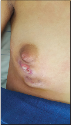 |
Figure 1: Idiopathic granulomatous mastitis. |
 |
Figure 2: MRI images breast revealing the presence of multiple breast collections from the right breast, fusing towards the axillary extension and towards the infero-external quadrant. |
Case 2
38-year-old patient, with no notable pathological history, consulting for non-infiltrated erosive erythematous-purplish plaques of the left breast, of different ages, slightly painful, evolving for 6 months, respecting the areolar region and the nipple, without nipple discharge or oozing (Fig. 3). Repeated biopsies performed on recent lesions showed a non specific inflammatory infiltrate without cyto-nuclear atypia, eliminating a neoplastic origin. Challenging treatments have been tried. First, antibiotic treatment, thinking of vegetative pyoderma, then very strong topical corticosteroids in the face of the possibility of contact eczema or pyoderma gangrenosum in its superficial form.
 |
Figure 3: Non specific chronic lesions evocating pathomimia. |
Despite a clinical evidence of self-inflicted injuries, the patient denied any involvement. The repeated questioning rediscovered the notion of family conflicts. The patient was hospitalized, and the lesions were covered with a dry bandage with a favorable evolution within a few weeks. The diagnosis of pathomimia was retained.
DISCUSSION
Idiopathic granulomatous mastitis is a rare benign condition. It mostly affects young women within 5 years of pregnancy or while breastfeeding. It always poses diagnostic problems, and constitutes a diagnosis of elimination [1].
This clinical aspect should lead to a number of diagnoses being discussed. Inflammatory breast cancer must be the fear of any such lesion. X-ray examinations should be done first to rule out this diagnosis, whether mammography in older women, or breast ultrasound and MRI in younger ones with predominantly glandular breasts [2].
Collecting pus is not always straightforward, as the high viscosity of the necrotic tissue sometimes does not allow aspiration. It is therefore sometimes necessary to carry out microbiological research on biopsy fragments. Bacteriological research should focus on aerobes and anaerobes, with a search for atypical germs. Molecular biology can be of great help in ruling out tuberculosis [3]. We can also suggest a pyoderma gangrenosum, or a diabetic mastopathy, hence the interest of the skin biopsy. In this case, after the histological study and bacteriological examinations, we are referred to the group of non-infectious granulomatous mastitis. History and paraclinical examinations should rule out the diagnosis of sarcoidosis, Wegner’s granulomatosis, or foreign body granuloma.
Pathomimia is defined as a deliberately self-induced factitious pathology without confusional or proven consciousness disorders [4]. Self-inflicted lesions are most often cutaneous, mimicking repeated infections or chronic wounds that are slow to heal. All areas of the body can be mutilated. The diagnosis of this disease is sometimes complex. The care is twofold: it requires psychological work associated with reinforced medico-surgical care [5].
CONCLUSION
Breast ulcerations are often sources of diagnostic difficulties, the reflex being to always think of an underlying mammary pathology, but certain purely dermatological conditions can advance the diagnosis.
Consent
The examination of the patient was conducted according to the principles of the Declaration of Helsinki.
The authors certify that they have obtained all appropriate patient consent forms, in which the patients gave their consent for images and other clinical information to be included in the journal. The patients understand that their names and initials will not be published and due effort will be made to conceal their identity, but that anonymity cannot be guaranteed.
REFERENCES
1. Boufettal H, Essodegui F, Noun M, Hermas S, Samouh N. Idiopathic granulomatous mastitis:a report of twenty cases. Diagn Interv Imaging. 2012;93:586-96.
2. Ennasser H, Raoudi JE, Taheri H, Saadi H, Mimouni A. [Idiopathic granulomatous mastitis:4 case-reports and literature review]. Pan Afr Med J. 2020;37:128.
3. Bouhout T, Serji B, Egyir EU, Amri BE, Bouhout I, Soufi M, et al. Breast tuberculosis:a case report. Pan Afr Med J. 2017;28:183.
4. Misery L. Cutaneous pathomimias. Ann Medico-Psycho. 2010;168:297–300.
5. Gieler U, Consolf S, Aragones L, Linder D. Self-inflicted lesions in dermatology:terminology and classification, a position paper from the European society for dermatology and psychiatry. Acta Derm Venereol. 2013;93:4–12.
Notes
Source of Support: Nil,
Conflict of Interest: None.
Request permissions
If you wish to reuse any or all of this article please use the e-mail (brzezoo77@yahoo.com) to contact with publisher.
| Related Articles | Search Authors in |
|
|
 http://orcid.org/0000-0002-2127-3863 http://orcid.org/0000-0001-7687-0158 http://orcid.org/0000-0002-2127-3863 http://orcid.org/0000-0001-7687-0158 |

Comments are closed.