Proliferating pilomatrixoma mimicking squamous cell carcinoma
Nuiakh Taoufik 1 , Naouar El Ouatassi1, Hamed Bamine1, Noureddine Mohammed El Alami El Amine1, Selma El Kadiri2, Hanane Baybay2, Zakia Douhi2, Sara Elloudi2, Fatima-Zahra Mernissi2, Nawal Hammas3, Layla Tahiri Elousrouti3, Hind El Fatemi3, Laila Chbani3
1Departement of Otorhinolaryngology, CHU Hassan II, Fez, Morocco, 2Department of Dermatology, CHU Hassan II, Fez, Morocco, 3Department of Pathology, Hassan II University Hospital, Fez, Morocco
Corresponding author: Nuiakh Taoufik, MD
Submission: 18.08.2020 Acceptance: 21.02.2021
DOI: 10.7241/ourd.2021e.4
Cite this article: Taoufi k N, El Ouatassi N, Bamine H, El Alami El Amine NM, El Kadiri S, Baybay H, Douhi Z, Elloudi S, Mernissi F-Z, Hammas N, Tahiri Elousrouti L, El Fatemi H, Chbani L. Proliferating pilomatrixoma mimicking squamous cell carcinoma. Our Dermatol Online. 2021;12(e):e4.
Citation tools:

Copyright information
© Our Dermatology Online 2021. No commercial re-use. See rights and permissions. Published by Our Dermatology Online.
Sir
Pilomatrixoma or calcifying epithelioma of Malherbe is an adnexal benign skin tumor arising from the hair follicle matrix cells. Although pilomatrixoma is a well-recognized lesion, it is frequently misdiagnosed as other tumors. Herein we report a case of a 45-year-old-woman of retro auricular pilomatrixoma mimicking squamous cell carcinoma.
A 45-year-old woman presented with a 6-months slowly growing retro auricular tumor. She didn’t report a history of trauma to the site. Dermatological examination revealed an ulcerated, lobulated and tumoral mass of 3-cm in diameter (Fig. 1). In front of this clinical presentation, we thought about many diagnoses such as squamous cell carcinoma, cutaneous metastasis, malignant pilomatricoma, and epidermal cyst. The tumor was removed with a 5-mm margin. Microscopic examination revealed hyperchromatic, pleomorphic, and basaloid cells with prominent nucleoli (Fig. 2). Centrally, it was composed of shadow cells (Fig. 3). The typical histologic pattern led to the diagnosis of proliferating pilomatricoma. Two years later, there was no evidence of either local recurrence or metastatic spread (Fig. 4).
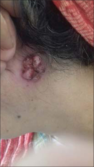 |
Figure 1: Clinical image showing an ulcerated, lobulated and tumoral mass. |
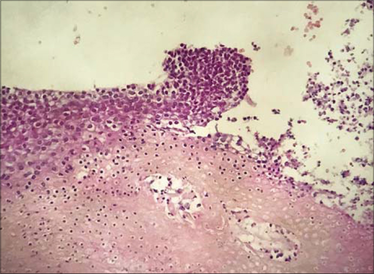 |
Figure 2: Microscopic examination revealed hyperchromatic, pleomorphic, and basaloid cells with prominent nucleoli. (HESx200). |
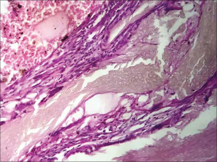 |
Figure 3: Microscopic examination showing central shadow cells (HESx200). |
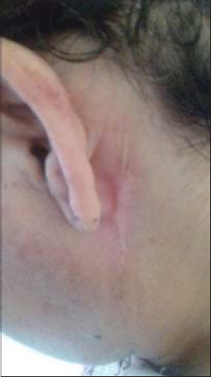 |
Figure 4: Re-examination 2 years later showing clinical remission of the disease with a significant scaring. |
Pilomatrixoma was first described by Malherbe Chenantais in 1880 as a benign tumor. Later, Forbis and Helwing demonstrated that the tumor arose from the hair follicle matrix cells with immunochemistry [1]. Typically, the clinical presentation is an asymptomatic, firm, slowly growing, and mobile nodule. The commonest location is the head and the neck area followed by the upper extremities, the trunk, and the lower extremities [2]. Proliferating pilomatrixoma is a rare variant of pilomatrixoma and it is characterized by a large and ulcerated nodule [3]. Histological examination reveals a higher number of mitotic figures implying that the increased number of basaloid calls within these lesions may be a function of the proliferation rate [4]. There are no features of lymphatic or perineural involvement. It is not necessarily known that proliferating pilomatrixoma is a precursor of malignant pilomatrixoma. Malignancy should be suspected if there is ulceration, asymmetrical borders, and local recurrence [1]. It is characterized bu large, asymmetrical, poorly circumscribed lesion of several basaloid aggregations with jagged borders and extension of matrical cells within the dermis. It may show of lymphatic or perineural invasion [5].
We herein reported a rare case of proliferating pilomatrixoma mimicking a malignant tumor. The final diagnosis should be made on an excision specimen only.
Consent
The examination of the patient was conducted according to the principles of the Declaration of Helsinki.
REFERENCES
1. Jones CD, Ho W, Robertson BF, Gunn E, Morley S. Pilomatrixoma. Am J Dermatopathol. 2018;40:631–41.
2. Cohen-Levy WB, Jose J, Rosenberg AE, Pretell-Mazzini J. Slowly growing mass on the left shoulder. Skeletal Radiology. 2017;47:117–8.
3. García-Montero P, Repiso-Jiménez JB, Fernández-Canedo I. Pilomatrixoma proliferante simulando malignidad. Actas Dermosifiliogr. 2018;109:359.
4. Sharma D, Agarwal S, Jain LS, Kamal V. Pilomatrixoma masquerading as metastatic adenocarcinoma. A diagnostic pitfall on cytology. J Clin Diagn Res. 2014;8:FD13–4.
Notes
Source of Support: Nil,
Conflict of Interest: None declared.
Request permissions
If you wish to reuse any or all of this article please use the e-mail (brzezoo77@yahoo.com) to contact with publisher.
| Related Articles | Search Authors in |
|
|
 http://orcid.org/0000-0003-3455-3810 http://orcid.org/0000-0003-3455-3810 |

Comments are closed.