Laugier–Hunziker syndrome: an exceptional cause of acquired hyperpigmentation
Siham Belmourida , Meriame Meziane, Nadia Ismaili, Laila Benzekri, Baderddine Hassam, Karima Senouci
Department of Dermatology-Venereology, Mohammed V University, IBN Sina Hospital, Rabat, Morocco
Corresponding author: Siham Belmourida, MD
Submission: 04.11.2020; Acceptance: 14.02.2021
DOI: 10.7241/ourd.2021e.27
Cite this article: Belmourida S, Meziane M, Ismaili N, Benzekri L, Hassam H, Senouci K. Laugier–Hunziker syndrome: an exceptional cause of acquired hyperpigmentation. Our Dermatol Online. 2021;12(e):e27.
Citation tools:

Copyright information
© Our Dermatology Online 2021. No commercial re-use. See rights and permissions. Published by Our Dermatology Online.
Sir,
Laugier–Hunziker syndrome (LHS) is an idiopathic macular hyperpigmentation of skin characterized by brownish black spots on oral mucosa including lips associated with longitudinal melanonychia of nails [1].
We present an original case of a young man presenting a generalized hyperpigmentation falling within the framework of (LHS).
28 years old, presented with history of generalized cutaneous pigmented spots that appeared 1 year earlier without any associated symptoms. On clinical examination, we found generalized hyperpigmented macules diffused all over the body (Figs. 1a – 1d) including the genitals, no suspicious lesion was found. The patient was in good health and was not taking any drug. He did not smoke and no skin lesion had preceded the appearance of these spots.
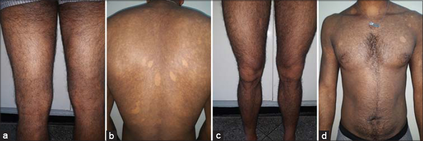 |
Figure 1: (a-d). Generalized hyperpigmented macules diffused all over the body. |
Although the lesions showed no signs of suspect and that the diagnosis of essential melanosis was evoked, we had performed a skin biopsy. Histopathological examination Showed that there was in places a discrete hyperpigmentation of the basal bed with migration melanin pigment in the superficial chorion where it is free or taken up by melanophages. There did not exist no melanocytic proliferation or associated lichenoid reaction. The aspect was therefore compatible with that of essential melanosis.
LHS was initially described in 1970 as an acquired, benign skin condition characterized by hyperpigmented macules on the lips and buccal mucosa associated with longitudinal melanonychia of nails [1,2]. Considered a diagnosis of exclusion and primarily reported from European countries in white population, sporadic cases have been reported from Asia including India in last decade. LHS occurs predominantly among middle-aged adults with a mean onset at 50 years of age and occurrence is usually seen after puberty. It is more prevalent in women and most reported cases have been in Whites, particularly in French and Italians. Reported cases have varied between second and ninth decades of life [2]. LHS is characterized by a varying number of asymptomatic, lenticular (lens-shaped), or linear, brown to black mucocutaneous macules, usually less than 5 mm in diameter. They may be single or confluent. They may have well-defined or indistinct margins. The hyperpigmentation occurs spontaneously and gradually and it is considered permanent [3]. The pathogenesis is thought to be linked to a functional alteration of the melanocytes that induces increased synthesis of melanosomes and subsequent transport to the basal cell layers. The etiology of the same is unknown. [2]. The pigmentary lesions carry no risk of malignant transformation. Cases have been reported in related family members. The condition has been variably reported as sporadic and inherited in an autosomal dominant fashion. Others have suggested that no genetic factors are associated with the syndrome [3]. Important differential diagnosis of LHS include Peutz Jeghers syndrome characterized by pigmentation around nose and mouth, oral cavity, palms, soles, and associated with hamartomatous gastrointestinal polyposis, which carries a high risk of malignancy. Addison’s disease, which is characterized by hyperpigmentation of the skin in areas subject to increased pressure, such as over the knuckles or the skin creases and in mucous membranes, is also an important differential diagnosis of LHS [2,4]. Treatment is sought mainly for cosmetic reasons and include Q-switched Nd: YAG or Q-switched alexandrite laser therapy for bothersome melanosis on the skin. Sun protection is important to prevent reoccurrence. Cryosurgery has also been tried for pigmentation in LHS with good results [5] for our patient, given that the lesions were generalized, and given the lack of means, he chose therapeutic abstention with sun protection.
Our case illustrates well the diversity of the geographical origin of the HS as what had been reported by the Indians and that it is not limited to a particular geographical region of the world
Consent
The examination of the patient was conducted according to the principles of the Declaration of Helsinki.
The authors certify that they have obtained all appropriate patient consent forms, in which the patients gave their consent for images and other clinical information to be included in the journal. The patients understand that their names and initials will not be published and due effort will be made to conceal their identity, but that anonymity cannot be guaranteed.
REFERENCES
1. Wei Z, Li GY, Ruan HH, Zhang L, Wang WM, Wang X. Laugier-Hunziker syndrome:A case report. J Stomatol Oral Maxillofac Surg. 2018;119:158-60.
2. Aboobacker Sha, Gunjan Gu. Laugier-Hunziker Syndrome. StatPearls. Treasure Island (FL);2020 Oct 13.
3. Duan N, Zhang YH, Wang WM, Wang X. Mystery behind labial and oral melanotic macules:Clinical, dermoscopic and pathological aspects of Laugier-Hunziker syndrome. World J Clin Cases. 2018;6:322-34.
4. MiličevićT, Žaja I, TešanovićD, Radman M. Laugier-Hunziker syndrome in endocrine clinical practice. Endocrinol Diabetes Metab Case Rep. 2018;2018:18-0025.
5. Abduljabbar T, Vohra F, Akram Z, Ghani SMA, Al-Hamoudi N, Javed F. Efficacy of surgical laser therapy in the management of oral pigmented lesions:A systematic review. J Photochem Photobiol B. 2017;173:353-9.
Notes
Source of Support: Nil,
Conflict of Interest: None declared.
Request permissions
If you wish to reuse any or all of this article please use the e-mail (brzezoo77@yahoo.com) to contact with publisher.
| Related Articles | Search Authors in |
|
|
 http://orcid.org/0000-0002-2795-9161 http://orcid.org/0000-0002-2795-9161 |

Comments are closed.