Trichilemmal carcinoma in the scalp: A case report
Asmae Abdelmouttalib 1, Achraf Khayri2, Mariame Meziane1, Karima Senouci1
1Dermatology and Venereology Department, Mohamed V University in Rabat, Morroco, 2Plastic Surgery Department, Mohamed V University in Rabat, Morroco
Corresponding author: Asmae Abdelmouttalib, MD
Submission: 25.11.2020 Acceptance: 12.02.2021
DOI: 10.7241/ourd.2021e.23
Cite this article: Abdelmouttalib A, Khayri A, Meziane M, Senouci K. Trichilemmal carcinoma in the scalp: A case report. Our Dermatol Online. 2020;12(e):e23.
Citation tools:

Copyright information
© Our Dermatology Online 2021. No commercial re-use. See rights and permissions. Published by Our Dermatology Online.
Sir,
Trichilemmal carcinoma (TC) is a rare malignant adnexal neoplasm with outer roots heath differentiation.TC is commonly located on the scalp, forehead and neck, trunk or the upper extremities. We report a case of trichilemmal carcinoma in the scalp, which might have been diagnosed as a squamous cell carcinoma but histopathological examination confirm a trichilemmal carcinoma.
A 77-year-old women presented at our institution with a mass on his scalp (Fig. 1). She reported that a mass had first been observed 4 years earlier and it gradually increased in volume with recurrent bleeding. On clinical examination, there was a budding tumor mass measuring 10 cm in diameter, painless and bleeding easily on contact. The histological study was in favor of a trichilemmal carcinoma of the cc with a trichilemmal-type keratinization noted at the level of tumor masses with an edematous stroma rich in vascular structures. A head-and-neck computed tomography (CT) scan showed no other pathological findings. The patient underwent tumor excision with margins of 1 cm (Fig. 2). The patient recovered fully and remained without complications or recurrences after a follow-up period of 24 months.
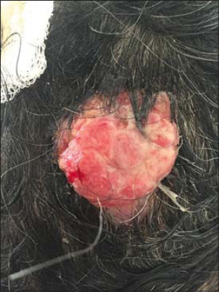 |
Figure 1: A budding tumor mass measuring, bleeding easily on contact. |
 |
Figure 2: Tumor excision with margins of 1 cm. |
Malignant hair follicle tumors account for 1% of adnexal carcinomas [1]. Trichilemmal carcinoma (TC) is a rare neoplasm that manifests as an asymptomatic exophytic or polypoid mass located in sun-exposed areas in older adults [2,3]. Clinical manifestations are variable, mimicking basal or squamous cell carcinomas, keratoacanthomas and nodular melanomas.TC usually behaves as a benign tumor and is amenable to curative resection with conventional surgery. Some authors suggest that safety margins greater than 1 cm are associated with increased overall survival [4]. Nonetheless, safety margins of 1 cm have been used safely and cost-effectively [5]. Additional therapeutic approaches include treatment with 5% imiquimod and adjuvant radiotherapy for patients with high-risk disease [2–6]. Since there are few articles reporting metastatic disease, magnetic resonance imaging and CT scan have been proposed as additional work-up studies for patients with TC. There are currently no guidelines for treatment or follow-up of TC, but it is recommended that these tumors be treated as non-melanoma skin cancers [2].
Trichilemmal carcinoma is a rare entity, which remains a challenge for dermatologists and pathologists. Examination of patients with trichilemmal cyst should take into account the possibility of malignant transformation. Close follow-up is necessary for early identification of recurrence.
Consent
The examination of the patient was conducted according to the principles of the Declaration of Helsinki.
The authors certify that they have obtained all appropriate patient consent forms, in which the patients gave their consent for images and other clinical information to be included in the journal. The patients understand that their names and initials will not be published and due effort will be made to conceal their identity, but that anonymity cannot be guaranteed.
REFERENCES
1. Sia PI, Figueira E, Allende A, Selva D. Malignant hair follicle tumors of the periorbital region:A review of literature and suggestion of a management guideline. Orbit. 2016;35:144-56.
2. Hamman MS, Brian Jiang SI. Management of trichilemmal carcinoma:an update and comprehensive review of the literature. Dermatol Surg. 2014;40:711-7.
3. Lee JH, Shin YW, Oh YH, Lee YJ. Trichilemmal Carcinoma of the Upper Eyelid:A Case Report. Korean J Ophthalmol. 2009;23:301-5.
4. Maya-Rico1 AM, Jaramillo-Pulgarín C, Londoño-García1 A, Peña-Zúñiga B. Locally aggressive trichilemmal carcinoma. An Bras Dermatol. 2018;93:579-81.
5. Zhuang SM, Zhang GH, Chen WK, Chen SW, Wang LP, Li H, et al. Survival study and clinicopathological evaluation of trichilemmal carcinoma. Mol Clin Oncol. 2013;1:499-502.
6. Chowdhry S, Jaiswal P, Dhali TK. Ulcerative giant solitary trichoepithelioma of scalp:a rare presentation. Our Dermatol Online. 2016;7:302-4.
Notes
Source of Support: Nil,
Conflict of Interest: None declared.
Request permissions
If you wish to reuse any or all of this article please use the e-mail (brzezoo77@yahoo.com) to contact with publisher.
| Related Articles | Search Authors in |
|
|
|

Comments are closed.