A rare case of Acute Febrile Neutrophilc Dermatosis – Sweet’s syndrome
Ashwini Kodigehalli Chikkanna Swamy , Sandhaya Prasad, Anjan Kumar Patra
Department of Dermatology, MVJ Medical College and Research Hospital Bangalore, India
Corresponding author: Ashwini Kodigehalli Chikkanna Swamy, MD
Submission: 03.08.2020; Acceptance: 09.01.2021
DOI: 10.7241/ourd.2021e.11
Cite this article: Swamy AKC, Prasad S, Patra AK. A rare case of acute febrile neutrophilc dermatosis – Sweet’s syndrome. Our Dermatol Online. 2021;12(e):e11.
Citation tools:

Copyright information
© Our Dermatology Online 2021. No commercial re-use. See rights and permissions. Published by Our Dermatology Online.
ABSTRACT
Sweet syndrome is an inflammatory dermatosis characterized by non-itchy, sometimes tender, erythematous plaques and papules most commonly distributed on the arms, upper body, head and neck. Histological findings include a dense dermal neutrophilic infiltrate with oedema. We report a case of 32year old female patient with solitary well defined erythematous edematous plaques with whitish scales at the centre of the lesion present over the extensor aspect of left forearm since 5days with sparing of palms , soles and mucosae. Laboratory investigations and histopathology were suggestive of Neutrophilic dermatoses (Sweet’s syndrome). This case was presented to highlight Atypical presentationof sweet syndrome – annular morphology and localization of lesions only over left forearm Therefore high index of suspicion is needed for its diagnosis in cases of solitary and isolated presentation along with necessary histopathological and laboratory investigations.
Key words: Sweet’s syndrome; Female; Dermal Neutrophilic dermatosis
INTRODUCTION
In 1964, R. D. Sweet published a description of eight women who presented with erythematous plaques, fever and non-specific infection .The exact etiology of Sweet syndrome is likely to be complex as numerous triggers can impact cell signaling pathways that lead to an increase in neutrophil production and migration into tissues [1].
Acute febrile neutrophilic dermatosis or Sweet syndrome is a condition characterized by fever, tender erythematous papules, nodules or plaques, neutrophilic leukocytosis, and dense dermal inflammatory infiltrates chiefly composed of polymorphonuclear neutrophils [2].
CASE REPORT
A 32 year old female patient came with chief complaints of red raised painful lesion over the left forearm since 5 days initially smaller in size gradually progressed to the present size within duration of 3-5 days. Patient also gave history of 2 episodes fever prior to the onset of lesion which was moderate grade intermittent not associated with chills and rigors. History of loose stools 2-3 episodes, blood tinged, non-foul smelling 1 week back for which she consulted a local doctor (medication details not known), became asymptomatic within 3days.
No h/o weight loss, loss of appetite, No h/o breathlessness, pain abdomen No h/o muscle weakness, No h/o bleeding per rectum or any other symptoms suggesting systemic disease.
No h/o traum, No h/o insect bite, No h/o loss of sensation over the lesion.
On General Physical Examination:
Vitals stable; Patient was febrile 100F; No lymphadenopathy/no pedal edema +
Systemic Examination: No abnormality detected.
On Cutaneous Examination: Two well defined erythematous edematous annular plaques measuring 6x 4cm with whitish scales present at the centre of the lesion present over the extensor aspect of lower 1/3 rd of left forearm . Margins of the scales were attached peripherally. Both Plaques showed similar morphology (Fig. 1) with sparing of palms and soles.
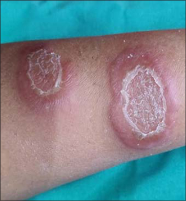 |
Figure 1: Two well defi ned erythematous edematous annular plaques measuring with whitish scales at the centre of the lesion present over the extensor aspect of lower 1/3 rd of left forearm. |
Sensation over the lesion was intact, No nerve thickening, Hair/Nail/Mucosa – Normal.
Clinically, Differential diagnosis of Sweet’s syndrome, Sarcoidosis, Jessner’s Lymphocytic infiltration, BT Hansen’s disease in type 1 reaction, was made.
Laboratory investigations are shown in the (Table 1) below revealed Leucocytosis with Neutrophilia, Raised C reactive protein and Raised ESR.
 |
Table 1: Shows Leucocytosis (Neutrophilia), Raised Erythrocyte sedimentation Rate Positive C-reactive protein |
KOH – Negative for fungal elements
Chest Xray – Normal
USG Abdomen and Pelvis – Normal
On Histopathological examination showed epidermis with mild acanthosis and spongiosis exocytosis of neutrophils into the epidermis. Dermis showed edema and dense neutrophilic infiltrate along with few lymphocytes and occasional eosinophils (Fig. 2). Also seen are perivascular and periadnexal inflammatory cell infiltrate chiefly composed of lymphocytes. No evidence of vasculitis present. Features were suggestive of Neurophilic Dermatoses – Sweet’s syndrome (Fig. 3).
 |
Figure 2: Histopathology showing mild acanthosis and spongiosis, exocytosis of neutrophils into the epidermis. Dermis showed edema and dense neutrophilic infi ltrate. |
 |
Figure 3: Undamaged vessel wall showing perivascular and periadnexal infl ammatory cell infi ltrate chiefl y composed of lymphocytes. |
Based on clinical and histopathological picture Diagnosis of Sweet’s Syndrome – Localised Neutrophilic Dermatosis was made. Patient was treated on Systemic steroids (Prednisolone 0.5mg/kg body weight) and topical steroids with emollients for 2weeks. Later patient was lost to follow up.
DISCUSSION
Sweet’s syndrome, or acute febrile neutrophilic dermatosis, is a rare inflammatory condition that is characterized by appearance of abrupt painful papulonodular skin lesion in the setting of a prodrome of fever, leukocytosis with neutrophilia, and pathological findings of neutrophilic infiltration of the upper dermis in the absence of Leukocytoclastic vasculitis. It is considered to be the major prototype of a subset of diseases known as neutrophilic dermatoses and is generally classified into 3 categories of classical (idiopathic), malignancy-associated and drug-induced Sweet’s syndrome, all of which share the same presenting scenario of abrupt onset of tender papulonodular skin lesions, most commonly affecting the face, neck, and upper extremities with asymmetrical distribution, in the setting of fever and leukocytosis, with histopathologic findings of dense neutrophilic infiltration of the dermis without evidence of vasculitis [3].
The classical Sweet’s syndrome is the most common type, which predominantly affects middle aged women and is usually associated with an infectious process, usually involving the upper respiratory or gastrointestinal tract, inflammatory bowel disease, or pregnancy [4].
The pathogenesis of Sweet’s syndrome remains unclear; however, the advances since its recognition have established the role of autoinflammatory processes involving both the innate and adaptive immune systems, eventually leading to their malfunction, resulting in immune-mediated hypersensitivity as well as involvement of cytokines such as interleukin1b (IL-1b), IL-17, and tumor necrosis factor-a (TNF-a) [4].
The classical Sweet’s syndrome is the most common type, which predominantly affects middle aged women and is usually associated with an infectious process, usually involving the upper respiratory or gastrointestinal tract, inflammatory bowel disease, or pregnancy. Skin lesion in cases associated with malignancy can be bullous or become ulcerated and resemble those of pyoderma gangrenosum [5]. The most common malignancies associated with Sweet’s syndrome are hematological malignancies, most commonly acute myelogenous leukemia. Solid tumors with carcinomas of the genitourinary tract, breast, and gastrointestinal tract have been reported as well. Drug-induced form of Sweet’s syndrome is most commonly observed with granulocyte-colony stimulating factor, all-trans retinoic acid, trimethoprim-sulfamthoxazole, and azathioprine [4,5].
Diagnosis of Sweet’s syndrome is based on typical clinical, laboratory and histological findings. Diagnostic criteria have been formulated to help clinicians to diagnose the disease. According to this criteria, Two major and Two minor criteria need to be fulfilled to make the diagnosis of Sweet syndrome [6].
Major Criteria
- Abrupt onset of painful erythematous plaques and nodules.
- Dense neutrophilic infiltrate without evidence of leukocytoclastic vasculitis by histopathology.
Minor Criteria
- Fever > 38 degree Celsius
- Association with an underlying haematologic or visceral malignancy, inflammatory disease, or pregnancy, or preceded by an upper respiratory or gastrointestinal infection or Vaccination
- Excellent response to treatment with systemic corticosteroids or potassium iodide.
- Abnormal laboratory values at presentation (three of four): erythrocyte sedimentation rate >20 mm/hour;positive C- reactive protein; > 8000 leucocytes; > 70% neutrophils.
The cutaneous manifestations persists for weeks or months if not treated. Although clinical signs and symptoms may spontaneously regress after treating the underlying cause , initial supportive treatment is generally required to control the disease. Mainstay of treatment is Corticosteroids.
Usual initial dose is 1mg/kg/day and tapered down over period of 4-6 weeks. Alternatives colchicine, potassium iodide, indomethacin, clofazimine, dapsone, Cyclosporin as secondary line treatment [6].
CONCLUSION
This case was presented to highlight Atypical presentation of sweet syndrome – Annular morphology and localization of lesions only over left forearm Therefore high index of suspicion is needed for its diagnosis in cases of solitary and isolated presentation along with necessary histopathological and laboratory investigations.
Consent
The examination of the patient was conducted according to the principles of the Declaration of Helsinki.
The authors certify that they have obtained all appropriate patient consent forms, in which the patients gave their consent for images and other clinical information to be included in the journal. The patients understand that their names and initials will not be published and due effort will be made to conceal their identity, but that anonymity cannot be guaranteed.
REFERENCES
1. Ormerod1 AD, Hampton PJ. Neutrophilic Dermatoses:Chapter 49 in Rook’s Textbook of Dermatology;9th edition (Vol 2). John Wiley &Sons Publisher;2016. pp 49.6–49.11.
2. Iqbal B, Kumar H, Kambale T. Acute febrile neutrophilic dermatosis (Sweet’s syndrome). Saudi J Health Sci. 2016;5:46-8.
3. Mollaeian A, Roudsari H, Talebi E. Sweet’s Syndrome:A classical presentation of a rare disease. J Invest Med High Impact Case Rep. 2019;7:1-5.
4. Villarreal-Villarreal C, Ocampo-Candiani J, Villarreal-Martínez A. Sweet syndrome:a review and update. Actas Dermosifiliogr. 2016;107:369-78.
5. Heath M, Ortega-Loayza A. Insights into the pathogenesis of Sweet’s syndrome. Front Immunol. 2019;10:414.
6. Khan U, Rizvi H, Ali F, Lebovic D. Sweet syndrome:a painful reality. BMJ Case Rep. 2016;2016:bcr 2016217606.
Notes
Source of Support: Nil,
Conflict of Interest: None declared.
Request permissions
If you wish to reuse any or all of this article please use the e-mail (brzezoo77@yahoo.com) to contact with publisher.
| Related Articles | Search Authors in |
|
|
|

Comments are closed.