Ovoid nests in nevocellular nevus
Soumaya Hamich1 , Basma Karrakchou1, Kaoutar Znati2, Karima Senouci1
1Department of Dermatology, Mohamed V University in Rabat, Ibn Sina University Hospital, Morocco, 2Department of Histopathology, Mohammed V University in Rabat, Ibn Sina University Hospital, Morocco
Corresponding author: Soumaya Hamich, MD
Submission: 11.07.2021; Acceptance: 17.10.2021
DOI: 10.7241/ourd.20223.20
Cite this article: Hamich S, Karrakchou B, Znati K, Senouci K. Ovoid nests in nevocellular nevus. Our Dermatol Online. 2022;13(3):315-316.
Citation tools:

Copyright information
© Our Dermatology Online 2022. No commercial re-use. See rights and permissions. Published by Our Dermatology Online.
We report the case of a 34-year-old female patient with no medical history presented herself with a lesion near the intergluteal cleft evolving for more than five years and gradually increasing in size. An examination revealed a painless nodule 1.5 × 2 cm in size, of soft consistency, and with a flesh-colored pedicle base with central hyperpigmentation and a depigmented peripheral area (Fig. 1). Dermoscopy revealed a hyperpigmented cerebriform structure in the center surmounted by several white scales and bordered by an irregular melanocytic network and multiple ovoid nests (Fig. 2).
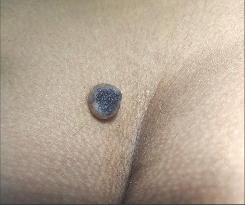 |
Figure 1: Nodule 1.5 × 2 cm in size with a flesh-colored pedicle base with central hyperpigmentation and a depigmented peripheral area. |
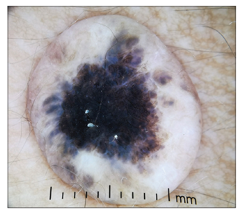 |
Figure 2: Dermoscopy of the lesion revealing a hyperpigmented cerebriform structure in the center and bordered by an irregular melanocytic network and multiple ovoid nests. |
Complete excision was performed. An anatomopathological examination revealed a tumor proliferation arranged in well-circumscribed epidermal and dermal nodules made of monomorphic polygonal cells with some mitotic figures (Fig. 3), suggesting a nevocellular nevus.
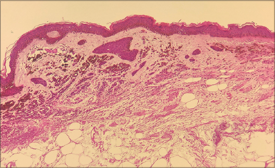 |
Figure 3: Histological image with a tumor proliferation arranged in well-circumscribed epidermal and dermal nodules made of monomorphic polygonal cells with some mitotic figures, suggesting a nevocellular nevus. |
In the present case, the lesion appeared similarly to basal cell carcinoma and seborrheic keratosis. On dermoscopic examination, it showed a cerebriform pattern and ovoid nests, suggesting these entities. These findings are not typically found in nevi. It is possible that this nevus was in its involution phase. Indeed, involutional or ancient nevi sometimes show signs of cellular atypia [1,2], which may be found by dermoscopy in ovoid nests.
In any case, it is essential to resort to a biopsy if in doubt to determine the exact nature of the tumor.
Consent
The examination of the patient was conducted according to the principles of the Declaration of Helsinki.
The authors certify that they have obtained all appropriate patient consent forms, in which the patients gave their consent for images and other clinical information to be included in the journal. The patients understand that their names and initials will not be published and due effort will be made to conceal their identity, but that anonymity cannot be guaranteed.
REFERENCES
1. Boyd AS, Chen SC, Shyr Y. Intradermal nevi with atypical nuclei in the elderly:The senescent nevus. J Am Acad Dermatol. 2015;73:500-6.
2. Kerl H, Wolf IH, Kerl K, Cerroni L, Kutzner H, Argenyi ZB. Ancient melanocytic nevus:A simulator of malignant melanoma. Am J Dermatopathol. 2011;33:127-30.
Notes
Source of Support: Nil,
Conflict of Interest: None declared.
Request permissions
If you wish to reuse any or all of this article please use the e-mail (brzezoo77@yahoo.com) to contact with publisher.
| Related Articles | Search Authors in |
|
|
|

Comments are closed.