Double-branched transungual acquired fibrokeratoma
Soumaya Hamich , Lina Mezni, Fatima Zahra El Gaitibi, Meriem Meziane, Karima Senouci
Department of Dermatology, University Hospital Center Ibn Sina, Rabat, Morocco
Corresponding author: Soumaya Hamich, MD
How to cite this article: Hamich S, Mezni L, El Gaitibi FZ, Meziane M, Senouci K. Double-branched transungual acquired fibrokeratoma. Our Dermatol Online. 2022;13(2):231-232.
Submission: 07.12.2020; Acceptance: 19.03.2021
DOI: 10.7241/ourd.20222.33
Citation tools:

Copyright information
© Our Dermatology Online 2022. No commercial re-use. See rights and permissions. Published by Our Dermatology Online.
Sir,
Acquired fibrokeratoma (AFK) is a rare benign skin tumor that usually manifests as a slow-growing solitary nodular lesion of the digit. It may appear on the fingers, toes, palms, or soles.
Herein, we report an atypical case of AK characterized by its transungual evolution and double-branched form.
A sixty-year-old male with a medical history of renal failure due to a chronic kidney disease of unknown etiology consulted for the evaluation of two lesions of the first right toenail evolving for the past one year.
The patient recalled a trauma one year before and the subsequent appearance of a cone at the proximal nail fold. Two months later, he noticed another cone emerging from the first. Both lesions were slow-growing and led to the consultation at our department.
A dermatological examination revealed two branching, longitudinal, and flesh-colored cones arising from the nail bed (Fig. 1), firm in consistency and with smooth surfaces on palpation.
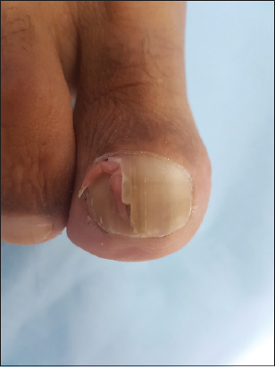 |
Figure 1: The two branching, longitudinal, and flesh-colored cones arising from the nail bed of the toenail. |
Keratotic materials were observed at the tips of the cones.
Both cones seemed to have the same place of emergence and were associated with a dissection of the nail plate. No pain or other symptoms were reported by the patient. A physical examination revealed no other lesions and there were no signs of tuberous sclerosis (Bourneville syndrome).
A dermoscopic examination found two filiform structures, 2 mm thick, emerging from the nail plate, with whitish-yellow and erythematous areas (Fig. 2).
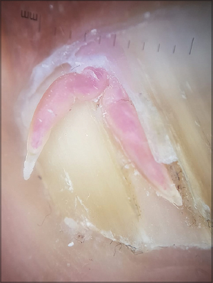 |
Figure 2: The two filiform structures, 2 mm thick, emerging from the nail plate with whitish-yellow and erythematous areas. |
We performed a complete excision of the lesions. After retracting the proximal nail fold, we found that the two cones were fused and their basal attachment was located under the proximal matrix (Fig. 3).
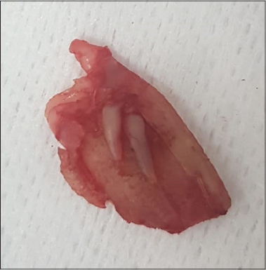 |
Figure 3: Surgical excision of the lesions, in which the two cones were fused and their basal attachment was located under the proximal matrix. |
Histologically, an acanthotic, papillomatous epidermis was present, surmounted by orthokeratotic hyperkeratosis. The dermis was characterized by fibrosis, thick-walled capillaries, and a mononuclear inflammatory infiltrate.
AF was first described in 1968 by Bart et al. [1], initially in the fingers but also in the palms, toes, and soles. Its etiology is presumed to be trauma or repetitive irritation, as in our case. Two cases were reported after a staphylococcal infection [2]. Qiao et al. reported a case of AK associated with cyclosporin use in a renal transplant patient. The increased presence of factor-XIIIa-positive dermal dendrocytes has also been suggested as a possible causal factor [1].
Hwang et al. classified the tumor into three subtypes according to the origin: periungual, subungual, and intraungual (or “dissecting” ungual) [3]. Urbina proposed the term transungual as another subtype, which fits more properly our case than intraungual [4] as the lesions in our patient did not develop through the whole thickness of the nail along the plate.
A rare variant known as multibranched acquired periungual fibrokeratoma (APF) was first described in a case report by Moriue et al. in 2014 [5]. Two more cases of multibranched APF were described by Goktay [6], who observed 2–5 mm thick multibranched structures protruding between the cuticle and the nail plate. To our knowledge, our observation is the first case of multibranched transungual acquired fibrokeratoma.
Owing to the diversity of vascular formations and collagen fiber accumulation, dermoscopic findings may often vary in different cases [1]. A central homogeneous pale-yellow area surrounded by a white, scaly, hyperkeratotic collarette was reported. Other authors described clumps of red homogenous lacunae divided by a white meshwork-like keratotic septum [7].
Histopathologic findings are hyperkeratosis, acanthosis, focal hypergranulosis of the epidermis, and the presence of thick bundles of collagen in the dermis with dilated capillaries oriented along the longitudinal axis of the tumor.
The main differential diagnosis is common warts, superficial acral fibromyxoma, a supernumerary digit, Koenen’s tumor, and cutaneous horns.
Acquired ungual fibrokeratomas do not regress spontaneously. The first-line treatment is surgical excision. The tumor has to be completely resected along with the basal attachment. Local recurrence is rare but may occur if a partial excision or curettage is performed.
Consent
The examination of the patient was conducted according to the principles of the Declaration of Helsinki.
The authors certify that they have obtained all appropriate patient consent forms, in which the patients gave their consent for images and other clinical information to be included in the journal. The patients understand that their names and initials will not be published and due effort will be made to conceal their identity, but that anonymity cannot be guaranteed.
REFERENCES
1. Shih S, Khachemoune A. Acquired digital fibrokeratoma:Review of its clinical and dermoscopic features and differential diagnosis. Int J Dermatol. 2019;58:151-8.
2. Amarouch H, Aitourghoui M, Ramli I, Zaouri H, Senouci K, Hassam B. [Post-staphylococcal acquired digital fibrokeratoma:A new case]. Presse Med. 2015;44:843-5.
3. Hwang S, Kim M, Cho BK, Park HJ. Clinical characteristics of acquired ungual fibrokeratoma. Indian J Dermatol Venereol Leprol. 2017;83:337–43.
4. Urbina F. Transungual fibrokeratoma. J Dermatol. 2017;44:e371.
5. Moriue T, Yoneda K, Moriue J, Nakai K, Kubota Y. Multibranched acquired periungual fibrokeratoma. JAMA Dermatol. 2014;150:456-7.
6. Göktay F, Altan ZM, Haras ZB, GüneşP, Yaşar Ş, Aytekin S, et al. Multibranched acquired periungual fibrokeratomas with confounding histopathologic findings resembling papillomavirus infection:A report of two cases. J Cutan Pathol. 2015;42:652-6.
7. Hayashi K, Matori S, Kariya Y, Sonosaki A, Yamaguchi S, Hagiwara K, et al. Dermoscopic observation of acquired digital fibrokeratoma developed on the dorsum of the fourth left toe. J Dermatol. 2016;43:107-8.
Notes
Source of Support: Nil,
Conflict of Interest: None declared.
Request permissions
If you wish to reuse any or all of this article please use the e-mail (brzezoo77@yahoo.com) to contact with publisher.
| Related Articles | Search Authors in |
|
|
|

Comments are closed.