Keloid-like dermatofibroma
Fatima Azzahra El Gaitibi 1, Soumaya Hamich1, Asmae Abdelmoutalib1, Kaoutar Znati2, Mariame Meziane1, Badereddine Hassam1, Karima Senouci1
1Department of Dermatology, Mohammed V University in Rabat, Ibn Sina University Hospital, Rabat, Morocco, 2Department of Histopathology, Mohammed V University in Rabat, Ibn Sina University Hospital, Rabat, Morocco
Corresponding author: Fatima Azzahra El Gaitibi, MD
How to cite this article: El Gaitibi FZ, Hamich S, Abdelmoutalib A, Znati K, Meziane M, Hassam B, Senouci K. Keloid-like dermatofibroma. Our Dermatol Online. 2021;12(4):460-461.
Submission: 31.01.2021; Acceptance: 16.04.2021
DOI: 10.7241/ourd.20214.27
Citation tools:

Copyright information
© Our Dermatology Online 2021. No commercial re-use. See rights and permissions. Published by Our Dermatology Online.
Dermatofibroma is a common benign skin tumor, mainly occurring in young to middle-aged females. It is frequently localized in the lower extremities. A typical dermatofibroma usually presents itself as a single firm papule or nodule, of variable color, bluish, brownish, or pinkish. Its clinical, dermoscopic, and histological features usually allow easy diagnosis [1]. However, it is possible to observe some variations of these typical features. Keloid-like dermatofibroma is one of these atypical presentations rarely reported in the literature [2].
A 40-year-old patient with no previous medical history presented to our dermatology department with a lumbosacral lesion evolving for several months. A physical examination revealed a firm, well-demarcated, asymptomatic erythematous nodule, 5 × 12 mm in size, localized in the lumbosacral area (Fig. 1). The patient denied any trauma preceding the onset of the lesion. There was no personal or familial history of keloidal scars. A dermoscopic examination revealed erythema, telangiectatic vessels, a shiny white streak, and a brownish-yellow pigmentation (Fig. 2). A biopsy was performed. A histological examination revealed an atrophic epidermis. The dermis contained a fibroblastic proliferation of low cell density haphazardly arranged, located on a fibromatous background (Figs. 3 and 4). Dermatofibroma with a keloidal presentation was the diagnosis.
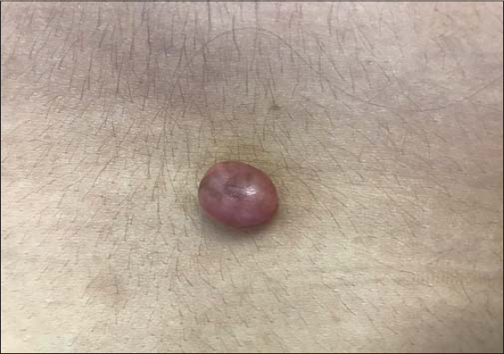 |
Figure 1: The firm erythematous nodule in the lumbosacral area. |
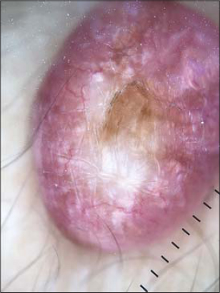 |
Figure 2: Erythema, telangiectatic vessels, a shiny white streak, and a brownish-yellow pigmentation. |
 |
Figure 3: An atrophic epidermis and a fibroblastic proliferation on a fibromatous background (H&E; 10×). |
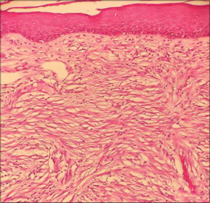 |
Figure 4: A fibroblastic proliferation of low cell density haphazardly arranged, located on a fibromatous background (H&E; 40×). |
In the present case, dermoscopic features were different from the signs usually found in dermatofibroma, which are a central white patch and a delicate pigment network. Indeed, a keloidal-like dermatofibroma presents itself as a pink nodule with other dermoscopic features. Erythema and vascular structures (arborizing vessels or/and dotted vessels) are the most frequently reported features. Besides, the shiny white streaks are present in around half of cases. These features may be suggestive of malignant lesions, especially amelanotic melanoma and basal cell carcinoma [3].
Consent
The examination of the patient was conducted according to the principles of the Declaration of Helsinki.
The authors certify that they have obtained all appropriate patient consent forms, in which the patients gave their consent for images and other clinical information to be included in the journal. The patients understand that their names and initials will not be published and due effort will be made to conceal their identity, but that anonymity cannot be guaranteed.
REFERENCES
1. Ben Rejeb S, Dhaoui A, Ben Ghachem D, Souissi A, Bellil K. Multiple eruptive dermatofibromas occuring in a patient under hemodialysis. Our Dermatol Online. 2016;7:412-4.
2. Kim JM, Cho HJ, Moon SH. Rare experience of keloidal dermatofibroma of forehead. Arch Craniofac Surg. 2018;19:72-4.
3. Llambrich A, Vila A, Terrasa F, Bañuls J, Nadal C, Zaballos P. Dermoscopy of pink nodular dermatofibromas:A study of 36 cases. Australas J Dermatol. 2019;60:e357-60.
Notes
Source of Support: Nil,
Conflict of Interest: None declared.
Request permissions
If you wish to reuse any or all of this article please use the e-mail (brzezoo77@yahoo.com) to contact with publisher.
| Related Articles | Search Authors in |
|
|
 http://orcid.org/0000-0002-0313-6152 http://orcid.org/0000-0002-0313-6152 |

Comments are closed.