Chronic keloidal nodules of the hand: Don’t forget the cutaneous leishmaniasis
Meryam Chaabani , Kahena Jaber, Faten Rebhi, Nejib Doss, Mohammed Raouf Dhaoui
Department of Dermatology, Military Hospital of Tunis, Tunisia
Corresponding author: Meryam Chaabani, MD
How to cite this article: Chaabani M, Jaber K, Rebhi F, Doss N, Dhaoui MR. Chronic keloidal nodules of the hand: Don’t forget the cutaneous leishmaniasis. Our Dermatol Online. 2021;12(3):324-325.
Submission: 09.01.2021; Acceptance: 29.04.2021
DOI: 10.7241/ourd.20213.22
Citation tools:

Copyright information
© Our Dermatology Online 2021. No commercial re-use. See rights and permissions. Published by Our Dermatology Online.
Cutaneous leishmaniasis (CL) remains endemic in certain regions of the world. Several atypical presentations have been described in the literature. Herein, we report an unusual case of CL due to Leishmania infantum (LI) manifesting itself as chronic keloidal nodules on the hand.
A 47-year-old patient from southeastern Tunisia presented at our department with non-healing nodules on the back of the right hand evolving for the last two years. The nodules recurred after surgical excision and antibiotic treatments. An examination revealed two non-ulcerated keloidal nodules on the back of the right hand, 2.5 cm and 1 cm in diameter (Fig. 1). A histopathological examination revealed hyperkeratosis, acanthosis, and dermal granulomatous inflammation with a predominantly histiocytic infiltrate containing small and spherical non-flagellated cells with bar-shaped paranuclear kinetoplasts. The diagnosis of chronic CL was retained. Species typing by polymerase chain reaction (PCR) revealed the presence of LI MON-24 in the skin lesions. The patient was treated with meglumine antimoniate intramuscularly with a good response.
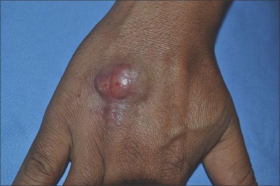 |
Figure 1: Non-ulcerated erythematous cutaneous nodules on the back of the right hand, without scales or crusts, 2.5 cm and 1 cm in size by the long axis. |
In Tunisia, CL is mainly caused by Leishmania major, LI, and Leishmania tropica (LT) [1]. Its clinical and progressive features depend on the infecting species and the immune response of the host [2]. Chronic CL (persistent for twelve months or more) is easily misdiagnosed because the lesions are often atypical and may be confused with other inflammatory and neoplastic skin diseases [3]. Furthermore, due to the low density of Leishmania bodies in this form, PCR is the most accurate diagnostic technique. The main causal agents of chronic CL are LT in the Old World and, less commonly, Leishmania braziliensis in the New World. Eroglu et al. reported LI to be a causal agent of the nodular form of chronic LC [3], as in our patient.
Consent
The examination of the patient was conducted according to the principles of the Declaration of Helsinki.
The authors certify that they have obtained all appropriate patient consent forms, in which the patients gave their consent for images and other clinical information to be included in the journal. The patients understand that their names and initials will not be published and due effort will be made to conceal their identity, but that anonymity cannot be guaranteed.
REFERENCES
1. Haouas N, Gorcii M, Chargui N, Aoun K, Bouratbine A, Messaadi Akrout F, et al. Leishmaniasis in central and southern Tunisia:Current geographical distribution of zymodemes. Parasite, EDP Scien. 2007;14:239-46.
2. Scott P, Novais FO. Cutaneous leishmaniasis:Immune responses in protection and pathogenesis. Nat Rev Immunol. 2016;16:581-92.
3. Eroglu F, Uzun S, Koltas IS. Comparison of clinical samples and methods in chronic cutaneous leishmaniasis. Am J Trop Med Hyg. 2014;91:895-900.
Notes
Source of Support: Nil,
Conflict of Interest: None declared.
Request permissions
If you wish to reuse any or all of this article please use the e-mail (brzezoo77@yahoo.com) to contact with publisher.
| Related Articles | Search Authors in |
|
|
|

Comments are closed.