Neglected mycosis fungoides transformed into cutaneous CD30¯ T-cell lymphoma
Mohamed El Amraoui 1, Naoufal Hjira1, Nadia Ismaili2, Mohammed Boui1, Karima Senouci2
1Department of Dermatology-Venereology, Mohammed V Military Training Hospital, Rabat, Morocco, 2Department of Dermatology-Venereology, Ibn Sina Hospital, Rabat, Morocco
Corresponding author: Dr. Mohamed El Amraoui
Submission: 22.12.2019; Acceptance: 23.02.2020
DOI: 10.7241/ourd.20211.26
Cite this article: El Amraoui M, Hjira N, Ismaili N, Boui M, Senouci K. Neglected mycosis fungoides transformed into cutaneous CD30? T-cell lymphoma. Our Dermatol Online. 2021;12(1):88-89.
Citation tools:

Copyright information
© Our Dermatology Online 2021. No commercial re-use. See rights and permissions. Published by Our Dermatology Online.
Sir,
Mycosis fungoides accounts for around half of primary cutaneous T-cell lymphomas. Prognosis is relatively favorable, but the possibility of several clinical presentations (large simulator) and the nonspecific nature of early eczema-like lesions delay the diagnosis and raise the possibility of transformation into other entities, whose prognosis might be more reserved and whose treatment might be intensified, as the following clinical case demonstrates [1–3].
A 45-year-old man with a history of chronic smoking, occasional alcoholism, and thin scaly arciform erythematous lesions evolving for more than 5 years and sitting at the roots of the limbs, buttocks, and sacral region (Fig. 1a and 1b). Consulted for an ulcerous, necrotic, and hemorrhagic tumor on the right elbow atop an arciform lesion, as much as 12 cm in diameter and evolving for 6 months in a context of conservation of the general condition (Fig. 2a). Clinical examinations revealed an axillary adenopathy 16 mm in diameter. Histology was in favor of mycosis fungoides with net epidermotropism without follicular mucinosis in the arcuate lesions. Mycosis fungoides transformed into CD30¯ large T-cell lymphoma as an elbow tumor with lymph node involvement. The tumor was staged as T3N2M0, and the patient received polychemotherapy (8CHOP) with a favorable evolution over five years of recoil (Fig. 2b and 2c). Our case highlights the particular advantage of keeping such diagnosis before any chronic dermatosis in adults and of knowing how to perform and repeat skin biopsies before any suspicion of dermatosis.
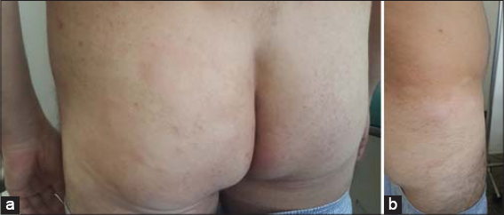 |
Figure 1: (a-b) Arcuate lesions with an infiltrated erythematous periphery and a hypopigmented center at the roots of the limbs, buttocks, and sacral region. |
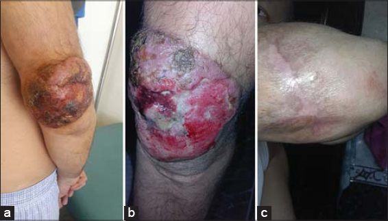 |
Figure 2: (a) An ulcero-budding tumor of the right elbow on a preexisting arciform lesion. (b-c) The tumor after the first and the last session of chemotherapy. |
Consent
The examination of the patient was conducted according to the principles of the Declaration of Helsinki.
The authors certify that they have obtained all appropriate patient consent forms, in which the patients have given consent for images and other clinical information to be included in the journal. The patients understand that their names and initials will not be published and due effort will be made to conceal their identity, but that anonymity cannot be guaranteed.
REFERENCES
1. Ichikawa S, Fukuhara N, Onishi Y, Ichinohasama R, Harigae H. Sustained remission of γδT-cell lymphoma by Graft-Versus-Lymphoma effect that relapsed early after cord blood transplantation. Clin Lymphoma Myeloma Leuk. 2018;18:e369-72.
2. Andruszkiewicz J, Brzezinski P. Mycosis fongoides mimic chronic eczema. Our Dermatol online. 2013;4;e3.
3. Mahalingam M, Reddy VB. Mycosis Fungoides, Then and Now…Have we travelled?Adv Anat Pathol. 2015;22:376-83.
Notes
Source of Support: Nil.
Conflict of Interest: None declared.
Request permissions
If you wish to reuse any or all of this article please use the e-mail (brzezoo77@yahoo.com) to contact with publisher.
| Related Articles | Search Authors in |
|
|
 http://orcid.org/0000-0001-7687-0158 http://orcid.org/0000-0001-7687-0158 |

Comments are closed.