Cutaneous side effects of hydroxyurea in a patient with thrombocytosis
Mirela Vasileva 1, Vesna Brishkoska Boshkovski2
1Clinical Hospital Stip, Stip, Republic of Macedonia, 2City Hospital “8th September” Skopje, Republic of Macedonia
Corresponding author: Dr. Mirela Vasileva
Submission: 16.10.2019; Acceptance: 08.01.2020
DOI: 10.7241/ourd.2020S3.3
Cite this article: Vasileva M, Brishkoska Boshkovski V. Cutaneous side effects of hydroxyurea in a patient with thrombocytosis. Our Dermatol Online. 2020;11(Supp. 3):10-12.
Citation tools:

Copyright information
© Our Dermatology Online 2020. No commercial re-use. See rights and permissions. Published by Our Dermatology Online.
ABSTRACT
Thrombocytosis is a disorder in which the body produces an abnormally high number of platelets. Hydroxyurea is an anticancer chemotherapy drug used for the treatment of cancer and thrombocytosis. Long-term use of hydroxyurea is associated with cutaneous adverse effects and complications such as leg ulcers and non-melanoma skin cancers. A sixty-year-old male with diagnosed thrombocytosis began treatment with oral hydroxyurea 1000 mg daily four years ago. A year ago, he developed leg ulcers. An examination revealed multiple ulcers the size of a metal pair on both legs, multiple actinic keratoses, and one SCC. We must be aware of the possibility that leg ulcers may be a complication of HU therapy as well as always be careful in taking medical histories. Every nonhealing wound should be completely explored for rapid diagnosis. Prevention and early intervention should, therefore, be the mainstay of treatment.
Key words: Trombocitosis; Hidroxyurea; Leg ulcer
INTRODUCTION
Thrombocytosis is a disorder in which the body produces an abnormally high number of platelets, which play an important role in blood clotting. Platelets are blood particles produced in the bone marrow. They quickly dissolve and their life lasts relatively briefly, on average from 3 to 5 days. Thrombocytosis may be caused by diseases of the blood and bone marrow. When thrombocytosis is caused by bone marrow disorders, it is called autonomic, primary, or essential thrombocytosis or essential thrombocythemia.
The treatment of reactive thrombocytosis is directed to the cause. If the cause is previous surgery or injury that caused significant blood loss, thrombocytosis will not last long.
Essential thrombocytosis, for its treatment, requires the use of cytostatics, antiviral drugs, and aspirin. In most cases, platelet levels return to normal after the treatment of the underlying cause.
Exceptionally, removing the spleen may cause lifelong thrombocytosis.
Hydroxyurea is an anticancer chemotherapy drug classified as an antimetabolite. It is used for the treatment of leukemia, thrombocytosis, head and neck cancers, malignant melanoma, and ovarian cancer in the case of no response to standard therapy. The side effects of hydroxyurea are very rare and almost always reversible after the completion of treatment.
The severity of the side effects depends on the dose of the drug: high doses more often cause unwanted effects.
The most common side effect is a low blood count, occurring in more than 30% of patients taking the drug.
The following side effects are less common: hair loss, nausea and vomiting, diarrhea, mouth sores, poor appetite, nail thickening, nail banding, discoloration of the skin or nails, darkening of the skin where a previous radiation treatment has been given.
In the last years, patients on long-term hydroxyurea and with cutaneous ulcerations have been reported.
Hydroxyurea and Skin Problems
Hydroxyurea is an oral antimetabolite that prevents DNA synthesis and promotes cell death by inhibiting ribonucleoside reductase, an important enzyme in the cell cycle. This drug is used in the treatment of chronic myeloproliferative neoplasms [1]. Longterm use of hydroxyurea is associated with cutaneous adverse effects and complications such as leg ulcers and nonmelanoma skin cancers [2].
Leg ulcers are a relatively frequent issue in patients with thrombocytosis under treatment with hydroxyurea. The pathogenesis may be multifactorial but remains unknown.
Concomitant arterial or venous disease may play a role in the occurrence of ulcerations.
CASE REPORT
We report a patient with diagnosed myeloproliferative disorders who developed hydroxyurea leg ulcers and multiple actinic keratoses. A sixty-year-old male was diagnosed with thrombocythemia five years ago. A year after, he began treatment with oral hydroxyurea 1000 mg daily. A year ago, he developed leg ulcers. An examination revealed multiple ulcers the size of a metal pair on both legs and multiple actinic keratoses. Because AK is difficult to differentiate from other malignancies with the naked eye, we made dermoscopy for the final diagnosis (Fig. 1). All the lesions were treated with cryotherapy except one, which was suspected for a squamous cell carcinoma. The suspicious lesion was removed surgically. Histopathological findings showed a moderately differentiated squamous cell carcinoma with a formation of keratin beads and mild chronic inflammation of the growth front and areas of infiltration (Figs. 2a and 2b).
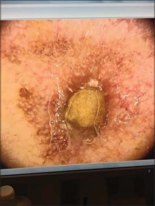 |
Figure 1: Dermoscopy of actinic keratosis growing into a squamous cell carcinoma: a central mass of keratin, a red pseudonetwork (“strawberry pattern”), and increasing neovascularization. |
 |
Figure 2: (a-b) Histopathological fi ndings showing a moderately differentiated squamous cell carcinoma with a formation of keratin beads and mild chronic infl ammation of the growth front and areas of infi ltration. |
The bilateral cutaneous ulcerations were painful and well-defined, with a livid border and a yellow fibrinous base. The ulcers had been open continuously without signs of healing for three months and had not responded to local treatment. The treatment involved compression bandages combined with a silver dressing and topical creams, gels, and ointments. None of these treatments gave the desired result.
Biopsy specimens were taken and histopathology revealed dermal fibrosis and occlusion of small capillaries.
Finally, we decided to replace the hydroxyurea with another drug in agreement with the hematologist.
Hydroxyurea treatment was discontinued and oral busulfan therapy was started. In just two months, we saw a reduction in the size of the ulcers (Figs. 3a and 3b), with compression bandages and an antiseptic dressing. In three months, the patient recovered completely (Figs. 4a and 4b).
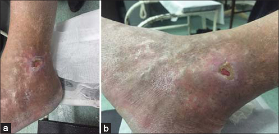 |
Figure 3: (a-b) Two months after discontinuation of therapy with hydroxyurea: well-defi ned with a livid border and a shallow base. |
 |
Figure 4: (a-b) Three months after discontinuation of therapy with hydroxyurea: hyperpigmentation at the site of the former ulcer. |
DISCUSSION
Hydroxyurea is usually well tolerated with a low toxic effect and is used for myeloproliferative disease therapy. However, cutaneous adverse effects, such as ulceration, have been described. Painful, nonhealing leg ulcers associated with hydroxyurea therapy have rarely been reported [3].
Numerous reported studies confirm the role of hydroxyurea therapy in the occurrence of leg ulcerations.
The pathogenesis of HU-induced ulceration may be multifactorial. Literature data reveals that most patients with HU-induced leg ulcers had been treated with more than 1 g of HU per day for at least one year [4,5]. Poor response to local and systemic therapy is a typical feature of HU-induced leg ulcers, and discontinuation of the drug is often required to achieve complete wound healing. In the case that we described, leg ulcerations developed after four years of treatment with HU. With the discontinuation of HU treatment, the ulcers were reduced. Sometimes, the management of complicated resistant HUrelated ulcers requires surgical therapy, such as skin grafting.
CONCLUSION
We must be aware of the possibility that leg ulcers may be a complication of HU therapy and always be extremely careful in taking medical histories. Complications may appear even years after HU treatment.
Every nonhealing wound should be completely explored for rapid diagnosis. Prevention and early intervention should, therefore, be the mainstay of treatment.
Consent
The examination of the patient was conducted according to the principles of the Declaration of Helsinki.
The authors certify that they have obtained all appropriate patient consent forms, in which the patients gave their consent for images and other clinical information to be included in the journal. The patients understand that their names and initials will not be published and due effort will be made to conceal their identity, but that anonymity cannot be guaranteed.
REFERENCES
1. Product Information. Hydroxyurea (hydroxyurea (hydroxyUREA)).“Par Pharmaceutical Inc, Chestnut Ridge, NY.
2. Antar A, Ishak RS, Otrock ZK, El-Majzoub N, Ghosn S, Mahfouz R, et al. Successful treatment of hydroxyurea-associated chronic leg ulcers associated with squamous cell carcinoma. Hematol Oncol Stem Cell Ther. 2014;7:166-9.
3. Thapa DP. Chemotherapy induced Beau’s and Mee’s line simultaneously:a case report and review of literature. Our Dermatol Online. 2019;10:77-8.
4. Fioramonti P, Fino P, Parisi P, Scuderi N, Onesti GM. A case of hydroxyurea-induced leg ulcer after definitive treatment suspension in a patient affected by thrombocythemia:effectiveness of a new collagenase. In Vivo. 2012;26:1053-6.
5. Ajili F, Mansour HB, Ghedira H, Zriba S, Metoui L, Gharsallah I, et al. Digital ischemia due to systemic sclerosis associated to essential thrombocythemia:A case report. Our Dermatol Online. 2013;4:508-10.
Notes
Source of Support: Nil.
Conflict of Interest: None declared.
Request permissions
If you wish to reuse any or all of this article please use the e-mail (brzezoo77@yahoo.com) to contact with publisher.
| Related Articles | Search Authors in |
|
|
 http://orcid.org/0000-0002-6807-5557 http://orcid.org/0000-0002-1580-6117 http://orcid.org/0000-0002-6807-5557 http://orcid.org/0000-0002-1580-6117 |

Comments are closed.