Foot pseudo-melanonychia
Patricia Chang 1, Marisol Gramajo2
1Department of Dermatology, Hospital General de Enfermedades IGSS and Hospital Ángeles, Guatemala; 2Department of Pathology, Hospital General de Enfermedades IGSS and Hospital Ángeles, Guatemala
Corresponding author: Dr. Patricia Chang
Submission: 20.02.2020; Acceptance: 02.05.2020
DOI: 10.7241/ourd.20203.18
Cite this article: Chang P, Gramajo M. Foot pseudo-melanonychia. Our Dermatol Online. 2020;11(3):305-306.
Citation tools:

Copyright information
© Our Dermatology Online 2020. No commercial re-use. See rights and permissions. Published by Our Dermatology Online.
A change in the color of the nails is referred to as nail dyschromia, discoloration of the nails, or chromonychia [1]. Black nail discoloration is called melanonychia.
We report a healthy 33-year-old female patient exhibiting onychopathy located in the nail plate of the third toe of the left foot consisting of a blackish coloration on the surface of and under the nail and on the second toe, slightly overlapping the third (Figs. 1a–1c). On dermatoscopy, the coloration is seen as blackish (Fig. 2a), and the microscope shows a linear, blackish-brown coloration (Fig. 2b). Scraping and cutting of the onychopathy are performed. Under the microscope, it is observed as a linear blackish-brown coloration, and the sample is refractive in polarized light (Fig. 2c). The surface of the nails after scraping and cutting can be seen in Figs. 3a–3b.
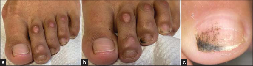 |
Figure 1: (a) Panoramic aspect of the lesion with an overlap of the second left ear. (b) Blackish coloration of the nail plate surface and underneath. (c) Dyschromia dermatoscopy of the nail plate surface. |
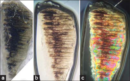 |
Figure 2: (a) Dermatoscopy of the nail plate sample. (b) Linear microscopy of the blackish lesion. (c) Birefringence of the nail with polarized light. |
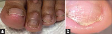 |
Figure 3: (a-b) Before and after nail curettage. |
The results of the rest of the physical exam fall within normal ranges. There is no significant family or personal history.
The patient reports that to have noticed the nail color change some time ago but cannot specify exactly when; she consults for fear of having nail melanoma.
A clinical diagnosis of pseudo-melanonychia onychopathy [2] was conducted favored by the overlap of the second toe.
Deformities of the toes can cause alterations in the nails due to the pressure that they bear and can manifest themselves in ways such as platonychia, frictional hematoma, frictional pseudo-hematoma, onycholysis, onychodystrophy, koilonychia, onychocryptosis, onychogryphosis, and pseudo-melanonychia [3].
Consent
The examination of the patient was conducted according to the principles of the Declaration of Helsinki.
The authors certify that they have obtained all appropriate patient consent forms, in which the patients have given consent for images and other clinical information to be included in the journal. The patients understand that their names and initials will not be published and due effort will be made to conceal their identity, but that anonymity cannot be guaranteed.
REFERENCES
1. Rubim A, Holzberg M, Baran R. Physical signs in:Baran &Dawbers Diseases of the nail and their management. Wiley Blackwell UK:2019:87-9.
2. Chang P. Pseudomelanoniquia podal asociada a clinodactilia. Dermatology DMCQ. 2013;11:158-9.
3. Chang P, Pinzón Porres P. Deformidades de los ortejos y alteraciones ungueales. Dermatology CMQ. 2008;6:232-40.
Notes
Source of Support: Nil,
Conflict of Interest: None declared.
Request permissions
If you wish to reuse any or all of this article please use the e-mail (brzezoo77@yahoo.com) to contact with publisher.
| Related Articles | Search Authors in |
|
|
|

Comments are closed.