An case report of gluteal erysipelas
Ibtissam Assenhaji , Hanane Baybay, Kaoutar Laamari, Asmae Rasso, Zakia Douhi, Sara Elloudi, Fatima Zahra Mernissi
Department of Dermatology, CHU Hassan II Fez, Morocco
Corresponding author: Dr. Ibtissam Assenhaji
Submission: 01.10.2019; Acceptance: 12.01.2020
DOI: 10.7241/ourd.20203.17
Cite this article: Assenhaji I, Baybay H, Laamari K, Rasso A, Douhi Z, Elloudi S, Mernissi FZ. An case report of gluteal erysipelas. Our Dermatol Online. 2020;11(3):303-304.
Citation tools:

Copyright information
© Our Dermatology Online 2020. No commercial re-use. See rights and permissions. Published by Our Dermatology Online.
Erysipelas is an acute infection of the upper dermis layer of the skin and the most frequent infection of soft tissues caused by group A β-hemolytic streptococci or Staphylococcus aureus [1]. Gluteal erysipelas is rarely described and not well characterized [2], reported in only 0.6% of all patients with erysipelas [3].
We report a case of a 61-year-old woman with no history of pathology or major surgery. A sudden development of a large area of erythema was observed in the left buttock. After two days, the erythema had spread to the abdomen and to the other buttock.
A general examination finds an apyretic patient in good condition, with a warm erythematous and edematous left buttock and half of the right buttock, and arriving at the left flank (Figs. 1 and 2). The size of the erythematous area was approx. 25–30 cm, and the border of the erythema was slightly elevated and clearly demarcated. Observed was also disseminated intergluteal (Fig. 3), submammary, inguinal, umbilical intertrigo and satellite lymphadenopathy. The rest of the somatic examination was unremarkable.
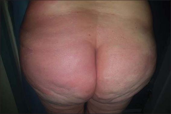 |
Figure 1: Warm erythematous and edematous left buttock and half of the right buttock. |
 |
Figure 2: Erythematous edema arriving at the left flank. |
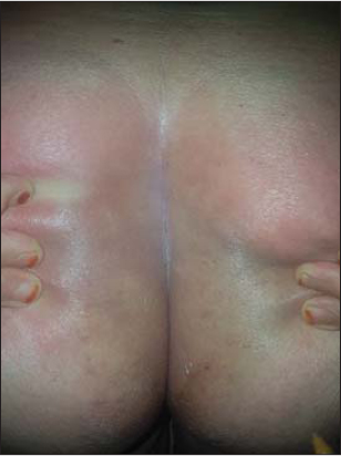 |
Figure 3: Intergluteal intertrigo. |
We clinically diagnosed these episodes as erysipelas because of the characteristic appearance of the erythema, even if this location, limited to the buttocks, is uncommon.
The patient’s white blood cell count was 9200/mL, and her C-reactive protein concentration was 81 mg/L. The erythema disappeared after 3 days of intravenous curative antibiotic therapy continued by oral antibiotics. The erysipelas was healed with the standardization of the infectious balance.
Consent
The examination of the patient was conducted according to the principles of the Declaration of Helsinki.
The authors certify that they have obtained all appropriate patient consent forms, in which the patients have given consent for images and other clinical information to be included in the journal. The patients understand that their names and initials will not be published and due effort will be made to conceal their identity, but that anonymity cannot be guaranteed.
REFERENCES
1. Aounallah A, Lahouel I, Belkahla M, Korbi M, Mbazaa A, Saidi W, et al. Erysipelas of the lower limb:Study of 400 cases. Our Dermatol Online. 2017;8(Suppl. 1):15-9.
2. Glatz M, Degen D, French LE, Aberer W, Müllegger RR. Erysipelas of the thigh and the gluteal region:retrospective multicenter analysis of a very rare entity in 39 patients. Dermatology. 2012;225:277-83.
3. Nagase T, Kadono T. Rare recurrent gluteal erysipelas associated with pressure ulcers in elderly patients:A report of two cases. Int J Gerontol. 2017;12:71-3.
Notes
Source of Support: Nil,
Conflict of Interest: None declared.
Request permissions
If you wish to reuse any or all of this article please use the e-mail (brzezoo77@yahoo.com) to contact with publisher.
| Related Articles | Search Authors in |
|
|
 http://orcid.org/000-0003-3455-3810 http://orcid.org/000-0003-3455-3810 |

Comments are closed.