A case of verrucous carcinoma of the back in a 17-year-old immunocompetent
Almamy Diabaté 1,2, Koffi Kouamé Pacôme Gbandama3, Julien Pitoni1, Mamadou Kaloga3, Christophe Bedane1
1Dermatology Department, Dupuytren Teaching Hospital, Limoges, France; 2Department of Dermatology, University Hospital of Bouaké, Ivory Coast; 3Department of Dermatology, University Hospital of Treichville, Abidjan, Ivory Coast
Corresponding author: Dr. Almamy Diabaté
Submission: 01.10.2019; Acceptance: 26.12.2019
DOI: 10.7241/ourd.20203.11
Cite this article: Diabaté A, Gbandama KKP, Pitoni J, Kaloga M, Bedane C. A case of verrucous carcinoma of the back in a 17-year-old immunocompetent. Our Dermatol Online. 2020;11(3):270-272.
Citation tools:

Copyright information
© Our Dermatology Online 2020. No commercial re-use. See rights and permissions. Published by Our Dermatology Online.
ABSTRACT
Verrucous carcinoma (CV) occurring in the oral cavity and in the genital area is induced by HPV. Our study describes a rare case of cutaneous CV manifested by a botiomyoma in a 17-year-old boy. Having no particular history, he consulted in dermatology at the Dupuytren Hospital in Limoges, for a tumor in the form of a nodular protrusion of angiomatous color in the lower back. Tumor biopsy and histological analysis revealed verrucous carcinoma. Our observation shows two originalities. The age of our patient is well below the average described by the literature as well as the unusual location of lesions. Verrucous carcinoma is a low-grade malignancy associated with HPV that can occur at any age and can be localized on all integuments.
Key words: Verrucous carcinoma, Adolescence, Back
INTRODUCTION
Verrucous carcinoma (VC) is a variant of well-differentiated squamous cell carcinoma and is considered to be associated with human papillomavirus (HPV) infection. Most VCs occur in the oral cavity, genital area or plantar surface [1,2]. VCs are rarely detected in the trunk area; the incidence of dermal VC from sites other than the inguinal region, buccal and leg is unknown. Surgical resection is the preferred treatment strategy for VC patients [1]. The present study describes a rare case of cutaneous VC of the back manifesting as a botiomyoma in a 17-year-old boy.
CASE REPORT
A 17-year-old sexually active heterosexual, nonsmoking, phototype IV male with no particular antecedent was sent to a dermatology consultation at Dupuytren Hospital in Limoges in November 2016 with a skin tumor in the lower back (Fig. 1). The patient reported that the lesion had appeared a month earlier with a painless papule and had gradually increased in size. On examination the tumor was in the form of a nodular lesion, budding angiomatous color, bleeding in places and measuring 6 cm in diameter. Anamnesis and clinical examination did not reveal suspicious lesions of papillomatosis or other changes. We discussed botriomycoma. Excisional biopsy of the lesion was performed, the histological examination of which showed a squamous mucosa with a papillomatous, acanthotic, hyperplastic, hyperkeratotic center with invagination of the keratin producing horny cysts. The dermis facing is very inflammatory seat of a dense infiltrate rich in lymphoplasmocytes, histiocytes and granulocytes. The epithelial basement membrane is nibbled by the inflammatory infiltrate and there are some casings of squamous cells isolated within the chorion, without basement membrane (Fig. 2). All these histological elements have concluded to warty squamous cell carcinoma. Complete patient assessment with abdominal ultrasound and MRI for abdominal cysts was negative. Serologies for hepatitis, HPV and HIV were also negative. The same is true for immunohistochemistry. The operative follow-up was uneventful and the patient healed well. Progress after 3 months postoperatively was without recurrence.
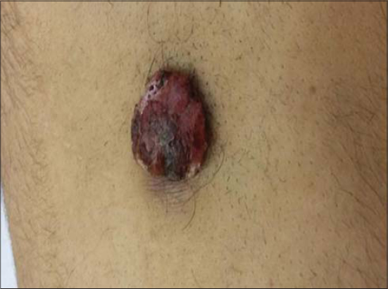 |
Figure 1: Lesion of verrucous carcinomas simulating botriomyoma. |
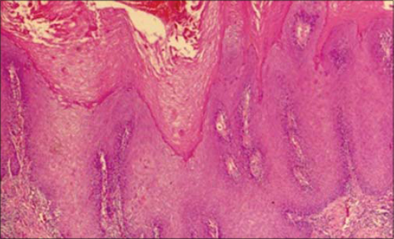 |
Figure 2: Histological picture of verrucous carcinoma. |
DISCUSSION
This observation draws its originality from two facts: occurrence in adolescence and localization on the back. Warty squamous cell carcinoma usually occurs in adulthood. Several authors report an age range between 63 and 91 years with a median age of 70 [2–5]. On the other hand, some authors have indicated 43 and 47 years in their case reports [1,6]. Our patient was only 17; such a young age is not reported in the literature. Male predominance has been found in the literature. According to the series of Terada [3] a verrucous lesion was observed in (70%) men and (30%) females. Similarly Kumar [2] mentioned a male predominance (94.7%). Our patient was male. In the majority of cases, the localization of verrucous carcinoma was the oral cavity, the genital area and the foot [1]. Some authors have described atypical localizations such as the neck, the armpits, the thigh and the buttocks in the fribro-epithelial polyp form [6–8]. Also, Terada [3] described 5 cases of VC of the skin in Japan. The sites were the hand, lip, face and foot sole. In our case, the lesion is located in the back. In addition, our patient underwent surgical resection without recurrence in a follow-up of three months. Surgical excision is the treatment of choice for small and well-defined lesions [1,3,7]. The effectiveness of several therapeutic is associated with the inclusion of radiotherapy, laser, immunotherapy, photodynamic therapy have been described in the literature [2,5]. The prognosis for recurrence of this tumor is controversial. According to Penera’s review of literature [1] recurrence occurred between 4 months and 3.5 years and also depended on the types of therapeutic measures used.
CONCLUSION
This study presents a rare case of cutaneous VC simulating a botriomycome appearing in the back, an unusual localization for this type of tumor and an exceptional finding in a young man of 17 years. Unlike VC lesions that occur at more common sites of development and in the elderly, HPV infection has not been identified in this case.
Consent
The examination of the patient was conducted according to the Declaration of Helsinki principles.
The authors certify that they have obtained all appropriate patient consent forms. In the form the patient(s) has/have given his/her/their consent for his/her/their images and other clinical information to be reported in the journal. The patients understand that their names and initials will not be published and due efforts will be made to conceal their identity, but anonymity cannot be guaranteed.
REFERENCES
1. Penera KE, Manji KA, Craig AB, Grootegoed RA, Leaming TR, Wirth GA. Atypical presentation of verrucous carcinoma:a case study and review of the literature. Foot Ankle Spec. 2013;6:318-22.
2. Paseiro Crespo G, Nebreda MG, BarcelóLópez M, Marqués Medina E, Gimeno Aranguez M. Verrucous carcinoma of the esophagus:A rare entity with a difficult diagnosis. Cir Esp. 2018;96:453-5.
3. Terada T. Verrucous carcinoma of the skin:a report on 5 Japanese cases. Ann Diagn Pathol. 2011;15:175-80.
4. Yuksel ME, Tamer F, Senyildiz Hasturk G. Umbilical squamous papilloma:A case report. Our Dermatol Online. 2019;10:408-9.
5. Terada T. Squamous cell carcinoma arising within verrucous carcinoma of the oral cavity:a case report. Int J Clin Exp Pathol. 2012;5:363-6.
6. Kurisu Y, Tsuji M, Yasuda E, Fujiwara M, Moriwaki S. Immunohistochemical findings and differential diagnosis of papillary-type cutaneous verrucous carcinoma of the neck:A case report. Oncol Lett. 2015;10:3823-5.
7. Nassiri A, Aqil N, Baybay H, Mernissi FZ, Souhli OA, Ahssaini M, et al. Extra genital HPV-6. Our Dermatol Online. 2019;10:71-3.
8. Helm MF, Haddad F, Farah R. What Is your diagnosis?Verrucous carcinoma. Cutis. 2015;96:82, 89-90.
Notes
Source of Support: Nil,
Conflict of Interest: None declared.
Request permissions
If you wish to reuse any or all of this article please use the e-mail (brzezoo77@yahoo.com) to contact with publisher.
| Related Articles | Search Authors in |
|
|
|

Comments are closed.