Clinical and dermoscopic features of a verrucous epidermal neavus
Sara Oukarfi , Rhizlane Chaoui, Salim Gallouj, Fatima Zahra Mernissi
Department of Dermatology CHU Hassan II, Fez, Morocco
Corresponding author: Dr. Sara Oukarfi, E-mail: oukarfisara@gmail.com
Submission: 30.07.2019; Acceptance: 18.12.2019
DOI: 10.7241/ourd.20202.29
Cite this article: Oukarfi S, Chaoui R, Gallouj S, Mernissi FZ. Clinical and dermoscopic features of a verrucous epidermal neavus. Our Dermatol Online. 2020;11(2):206-207.
Citation tools:

Copyright information
© Our Dermatology Online 2020. No commercial re-use. See rights and permissions. Published by Our Dermatology Online.
Sir
Epidermal nevi are defined as cutaneous hamartomas with several clinical forms. The verrucous form is the typical and most frequent form of epidermal neavus. We presented a 4 years old boy, with no particular medical history, was admitted for linear pigmented lesions of the right upper limb evolving since birth. Dermatological examination unveiled linear pigmented plaques starting at the dorsal surface of the thumb reaching to the elbow, well defined with irregular contours, with warty surface, resting on a skin hypopigmented in places (Fig. 1).
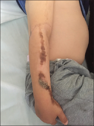 |
Figure 1: Clinical image of verrucous epidermal neavus. |
Dermoscopy revealed a cerebral aspect of the lesions, brown dots, white and brown exophytic papillary structures, and thick, adherent scales with some dot vessels.
The diagnosis was a verrucous epidermal neavus (VEN) and a CO2 laser was proposed as a treatment (Figs. 2 – 4).
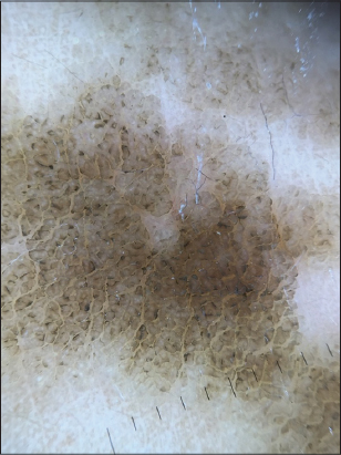 |
Figure 2: Thick branched brown lines and Cerebriform structures. |
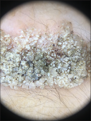 |
Figure 3: Thick adherent scales, dot vessels, papilliforme aspect. |
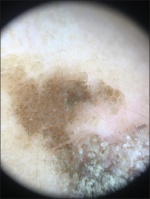 |
Figure 4: Cerebriform structures. |
The dermoscopic aspects of VEN have been described in a few articles; two studies were the subject of these aspects by Carbotti et al. [1] analyzing 8 patients with VEN, and more recently Elmas and Akdeniz [2] who treated 20 cases.
Carbotti et al. [1] reported lesions of VEN showed large brown circles and the absence of cysts, pigmented networks, and globules similar to seborrheic keratoses.
Elmas and Akdeniz [2] identified new non-vascular dermoscopic aspects that had not previously been described for VEN. These findings are thick branched brown lines, terminal hairs, brown dots, white and brown exophytic papillary structures, thick and adherent thick scales, thick branched lines, brown dots, brown serpiginous cerebral structures. With regard to the dermoscopic description of the vessels, the common aspect was the point vessels; the coiled, looped, serpiginous and polymorphic vessels were also present in some lesions.
In conclusion, verrucous epidermal neavuses may have dermoscopic characteristics similar to those of seborrheic keratosis and dermal nevi.
We suggest that dermoscopic examination may help in the diagnosis of VEN with decreased biopsy use. However, if there is a vascular appearance, polymorphic, which may be a malignant growth index, it would be reasonable to confirm the diagnosis by histopathology.
Consent
The examination of the patient was conducted according to the Declaration of Helsinki principles.
REFERENCES
1. Carbotti M, Coppola R, Graziano A, Verona Rinati M, Paolilli FL, et al. Dermoscopy of verrucous epidermal nevus:large brown circles as a novel feature for diagnosis. Int J Dermatol. 2016;55:653-6.
2. Elmas ÖF, Akdeniz N. Dermoscopic aspect of verrucous epidermal nevi:new findings. Turk J Med Sci. 2019;49:710-4.
Notes
Source of Support: Nil.
Conflict of Interest: None declared.
Request permissions
If you wish to reuse any or all of this article please use the e-mail (brzezoo77@yahoo.com) to contact with publisher.
| Related Articles | Search Authors in |
|
|
 http://orcid.org/0000-0001-7687-0158 http://orcid.org/0000-0001-7687-0158 |

Comments are closed.