Rosettes within rosacea
Niema Aqil , Aicha Nassiri, Kaoutar Moustaide, Salim Gallouj, Fatima Zahra Mernissi
Department of dermatology, Hassan II University Hospital Center, Fes, Morocco
Corresponding author: Dr. Niema Aqil, E-mail: niemaaqil90@gmail.com
Submission: 24.06.2018; Acceptance: 29.10.2018
DOI: 10.7241/ourd.20193.29
Cite this article: Aqil N, Nassiri A, Moustaide K, Gallouj S, Mernissi FZ. Rosettes within rosacea. Our Dermatol Online. 2019;10(3):314
Citation tools:
BibTex | CSV | RIS | refer/BiblX | Endnote XML | Wikipedia Citation Templates
Copyright information
© Our Dermatology Online 2019. No commercial re-use. See rights and permissions. Published by Our Dermatology Online.
Sir,
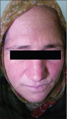
A 43-year-old woman presented with recurrent flushing of the face and redness, erythematous lesions of the face with hypersensitivity to heat. These symptoms had persisted for 4 years, with intermittent remissions lasting up to 2 months. A physical examination revealed facial erythema, telangiectasia, papules and pustules of the midfacial region with some scales and crusts. (Fig. 1) Dermoscopy revealed linear vessels characteristically arranged in a polygonal network, creamy and whitish linear areas and a clear rosette sign. (Fig. 2) The rest of the somatic examination was without abnormalities. An ophthalmological examination showed no evidence of keratitis, conjunctivitis or blepharitis. On these bases a diagnosis of papulopustular rosacea was made. The patient was treated with doxycycline for a total of 12 weeks, which led to a significant improvement.
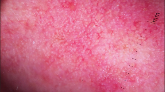
The rosette sign has been previously observed by Cuellar and colleagues and has been described as a new dermoscopic sign in actinic keratoses, which may be due to alternating areas of orthokeratosis and parakeratosis [1]. Recently, Liebman and his collaborators have pointed out that the rosette sign is an optical effect of polarized light and that its interaction with keratin-filled adnexal openings is observable in a wide range of cutaneous neoplasia [2]. This correlation could also explain the presence of this sign in rosacea too.
Consent
The examination of the patient was conducted according to the Declaration of Helsinki principles.
REFERENCES
1. Cuellar F, Vilalta A, Puig S, Palou J, Salerni G, Malvehy J. New dermoscopic pattern in actinic keratosis and related conditions. Arch Dermatol. 2009;145:732.
2. Liebman TN, Scope A, Rabinovitz H, Braun RP, Marghoob AA. Rosettes may be observed in a range of conditions. Arch Dermatol. 2011;147:1468.
Notes
Source of Support: Nil
Conflict of Interest: None declared.
Request permissions
If you wish to reuse any or all of this article please use the e-mail (brzezoo77@yahoo.com) to contact with publisher.
| Related Articles | Search Authors in |
|
|
|

Comments are closed.