Melanonychia in children
Ouiame EL Jouari , Ghita Senhaji, Salim Gallouj, Hanane Baybay, Fatima Zahra Mernissi
Department of Dermatology, University Hospital Hassan II Fez, Morocco
Corresponding author: Dr. Ouiame EL Jouari, E-mail: eljouariouiame@gmail.com
Submission: 14.09.2018; Acceptance: 04.12.2018
DOI: 10.7241/ourd.20193.23
Cite this article: EL Jouari O, Senhaji G, Gallouj G, Baybay H, Mernissi FZ. Melanonychia in children. Our Dermatol Online. 2019;10(3):302-303.
Citation tools:
BibTex | CSV | RIS | refer/BiblX | Endnote XML | Wikipedia Citation Templates
Copyright information
© Our Dermatology Online 2019. No commercial re-use. See rights and permissions. Published by Our Dermatology Online.
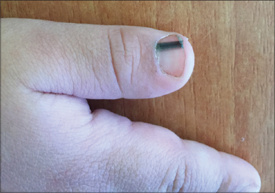
A healthy 2 years-old-boy, with no family history of melanoma or other tumors. He was brought in consultation by his mother for evaluation of a congenital lesion of the nail of the thumb of the left hand. Physical examination showed a homogeneous erythematous-brown color band of the nail of the thumb of the left hand. Without hyperpigmentation of the proximal nail fold (Fig. 1). Dermoscopy revealed multiple homogeneous, narrow and regular longitudinal lines in the nail plate with some pigmented globules. Without periungual pigmentation (Fig. 2). Correlation of the age, the congenital onset, clinical and dermoscopic findings suggested melanocytic nevus of the nail matrix as the most likely diagnosis. Given the apparent benign nature of the lesion, close clinical and dermoscopic control was recommended. After 24 months of follow-up, the size of the lesion was unchanged.
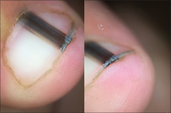
Melanonychia is defined as a brown or black pigmentation on a nail as a result of melanin and other exogenous pigments [1]. In rare instances, congenital nevi present subungually, with fewer than 20 biopsy-proven cases reported [2]. Nail matrix nevus usually presents with longitudinal melanonychia, which is also observed in subungual melanoma or other benign conditions such as subungual lentigo. Pigment bands of Nail matrix nevus are well demarcated from adjacent uninvolved nail plate devoid of pigment and the nail plate surrounding the longitudinal melanonychia also presents with a background of brown pigmentation [3]. Hutchinson sign is defined by longitudinal melanonychia extending into the periungual tissues. Whereas it is one criterion for the ABC rule of subungual melanoma and is traditionally considered a worrisome feature, it is certainly not pathognomonic and a malignant cause should not be assumed without thorough assessment [2]. Dermoscopy is especially helpful in the differential diagnosis of nail pigmentations, because it can reduce unnecessary and invasive nail unit biopsy. It usually observed as a limited width of brown background with a regular pattern of the longitudinal lines. A previous dermoscopic study of adult and childhood NMNs found that pseudo-Hutchinson’s sign, triangle sign, and globules were more frequent in childhood NMN and that Hutchinson’s sign and irregular band pattern also tended to be more common in children [1]. The management of pigmented nail lesions will therefore depend on whether the rare but very serious childhood melanoma is suspected. In cases of melanonychia in which malignancy is suspected (a broad band of pigment, Hutchinson sign, irregular dermoscopic features, a dark-skinned patient), the lesion must be completely excised. Dermoscopic and clinical follow-up should be reserved for lesions with low-risk features (narrow bands, uniform dermoscopic characteristics, no changes overtime) [3].
Consent
The examination of the patient was conducted according to the Declaration of Helsinki principles.
REFERENCES
1. Lee JH, Lim Y, Park JH, Lee JH, Jang KT, Kwon EJ, Clinicopathologic features of 28 cases of nail matrix nevi (NMNs) in Asians: Comparison between children and adultsJ Am Acad Dermatol 2018; 78: 479-89.
2. Goldminz AM, Wolpowitz D, Gottlieb AB, Krathen MS, Congenital subungual melanocytic nevus with a pseudo-Hutchinson signDermatol Online J 2013; 19: 8-
3. Agusti-Mejias A, Messeguerb F, Febrera I, Alegre V, Nevus melanocítico congénito subungueal y periunguealActas Dermosifiliogr 2013; 104: 446-8.
Notes
Source of Support: Nil
Conflict of Interest: None declared.
Request permissions
If you wish to reuse any or all of this article please use the e-mail (brzezoo77@yahoo.com) to contact with publisher.
| Related Articles | Search Authors in |
|
|
 http://orcid.org/000-0003-3455-3810 http://orcid.org/000-0003-3455-3810
|

Comments are closed.