Epidermal cysts of scrotum
Ouiame El Jouari 1, Anas Zaougui2, Ghita Senhaji1, Salim Gallouj1, Moulay Hassan Farih2, Fatima Zahra Mernissi1
1Department of Dermatology, University Hospital Hassan II, Fez, Morocco; 2Department of Urology, University Hospital Hassan II Fez, Morocco
Corresponding author: Dr. Ouiame El Jouari, E-mail: eljouariouiame@gmail.com
Submission: 27.05.2018; Acceptance: 24.09.2018
DOI:10.7241/ourd.20192.23
Cite this article: El Jouari O, Zaougui A, Gallouj S, Farih MH, Mernissi FM. Epidermal cysts of scrotum. Our Dermatol Online. 2019;10(2):197-197.
Citation tools:
BibTex | CSV | RIS | refer/BiblX | Endnote XML | Wikipedia Citation Templates
Copyright information
© Our Dermatology Online 2019. No commercial re-use. See rights and permissions. Published by Our Dermatology Online.
A 29-year-old man, without significant pathological antecedents. He consulted for asymptomatic scrotal papules, evolving for 5 months, gradually increasing in size and number. Clinical examination had found multiple skin color papules and nodules, firm in consistency, painless, arising from scrotal skin (Figs. 1a and 1b). A biopsy excision of a scrotal nodule was executed. The pathological study revealed an epidermoid cyst.
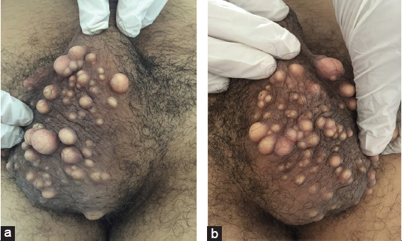
Epidermal cysts are benign epithelial cysts. In most cases, epidermal cysts occur in the skin of the scalp, ear, face, back and rarely scrotum. They consist of a sac lined by stratified squamous epithelium filled with laminated keratin, cholesterol crystals and debris. The main differential diagnosis is scrotal calcinosis. Treatment consists of a complete excision of the cyst to prevent recurrence.
Notes
Source of Support: Nil
Conflict of Interest: None declared.
Request permissions
If you wish to reuse any or all of this article please use the e-mail (brzezoo77@yahoo.com) to contact with publisher.
| Related Articles | Search Authors in |
|
|
|

Comments are closed.