|
Get Citation
|
|
|
Malakar S, Pal A, Pradhan P. Palisade pigmentation – describing a new dermoscopic fi nding. Our Dermatol Online. 2018;10(1):106-107. |
|
|
Download citation file:
|
Palisade pigmentation – describing a new dermoscopic finding
Subrata Malakar, Anjali Pal, Pratibha Pradhan
Rita Skin Foundation, Kolkata, India
Corresponding author: Dr. Anjali Pal, E-mail: unjelly@gmail.com
Submission: 24.04.2018; Acceptance: 19.06.2018
DOI:10.7241/ourd.20191.32
Sir,
Discoid lupus erythematosus (DLE) represents the most common subtype of cutaneous lupus erythematosus. There is a morphological diversity seen in DLE, at different stages of progression of the disease. Based on these features, dermoscopic criteria have been formed for both early and late stage DLE, and their histopathological correlations studied [1]. We would like to describe a new dermoscopic feature that we have observed in late stage DLE- palisade pigmentation.
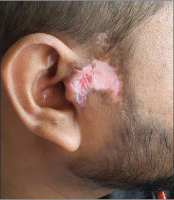
Case 1: A 26year old male presented with a lesion over the right pre-auricular area, of nine months duration. Examination revealed an irregularly shaped, depigmented atrophied plaque with a central erythematous area and overlying adherent scales, hyperpigmented border, and absence of hair follicles (Fig. 1). Dermoscopy revealed white shiny structures, telangiectasia, white scales, and palisade pigmentation (Fig. 2).

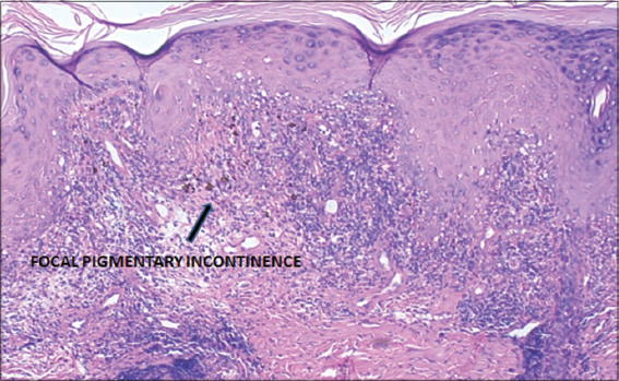
A biopsy from the lesion showed features confirming the diagnosis of DLE (Fig. 3).
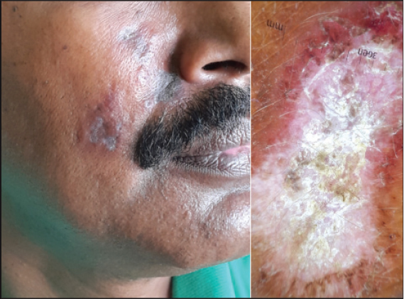
Case 2: A 35year old man noticed multiple lesions on his right cheek, over the past six months. They were asymptomatic. Cutaneous examination revealed multiple irregular violaceous atrophic plaques on the right cheek, with an erythematous border. Dermoscopy revealed white shiny structures, thick white overlying scales, telangiectasia, and palisade pigmentation (Fig. 4). Biopsy confirmed the diagnosis of DLE.
A ‘palisade’ is a fence of wooden stakes or iron railings fixed in the ground, forming an enclosure or defence. In dermatopathology, the term has been used to describe the typical arrangement of basaloid cells in basal cell carcinoma, and in granuloma annulare, for histiocytes surrounding an area of altered collagen [2].
‘Palisade pigmentation’ observed on dermoscopy here, is formed by the arrangement of pigment structures at the periphery of the lesion in late stage DLE. It appears as linear hyperpigmented streaks. They may be present focally or segmentally, and may extend for a distance, outwards, from the periphery of the lesion. Histopathologically, it corresponds to pigmentary incontinence, which was confirmed in our case. The incontinence may not involve the entire dermis, but is present focally or segmentally, and is due to focal interface vacuolar degeneration of the basal layer.
Hyperpigmentation has been described in dermoscopy of late stage DLE [1,3] in the form of honeycomb network, perifollicular pigmentation, radial pigment streaks or pigmentation arranged in unspecified patterns. In a study by Lallas et al [1], hyperpigmentation was observed on dermoscopy in 43.6% cases of DLE, and pigmentary incontinence was seen in 49.1% of the corresponding biopsies.
We would like to suggest this characteristic finding of ‘palisade pigmentation’ as a dermoscopic indicator of late stage DLE. Recognition of this pattern, in conjunction with other already established dermoscopic features, can help clinch the diagnosis.
CONSENT
The examination of the patient was conducted according to the Declaration of Helsinki principles.
REFERENCES
1. Lallas A, Apalla Z, Lefaki I, Sotiriou E, Lazaridou E, Ioannides D, et al. Dermoscopy of discoid lupus erythematosus. Br J Dermatol. 2013;168:284-8.
2. Madke B, Chougule BD, Kar S, Khopkar U. Appearances in clinical dermatology. Indian J Dermatol Venereol Leprol. 2014;80:432-47.
3. Errichetti E, Stinco G. Dermoscopy in General Dermatology:A Practical Overview. Dermatol Ther. 2016;6:471-507.
Notes
Source of Support: Nil
Conflict of Interest: None declared.
Comments are closed.