|
Get Citation
|
|
|
Madhumitha M, Sundaramoorthy S. A case of vitiligo with hypertrophic lichen planus – an autoimmune association. Our Dermatol Online. 2018;9(4):437-439. |
|
|
Download citation file:
|
A case of vitiligo with hypertrophic lichen planus – an autoimmune association
Mohanraj Madhumitha, Srinivasan Sundaramoorthy
Chettinad Hospital and Research Institute, Kelambakkam, Tamil Nadu, India
Corresponding author: Prof. Srinivasan Sundaramoorthy, E-mail: hamsrini@yahoo.co.in
Submission: 01.02.2018; Acceptance: 18.04.2018
DOI: 10.7241/ourd.20184.21
ABSTRACT
Vitiligo is a common autoimmune dermatological disorder characterized by depigmentation of the skin and mucous membranes. It has been associated with a variety of autoimmune disorders. A 54 years old male patient presented with depigmented patches over the legs for 30 years and itchy hyperpigmented plaques over the forehead for past 2 months. Patient had history of recurrent oral ulcers and history of psychological stress. Cutaneous examination revealed two well defined depigmented patches over the shin of both legs, and over lower lip. There were multiple hyperpigmented violaceous plaques over the forehead, right side cheek and scalp. White streaks were present over the buccal mucosa. Biopsy report for the hyperpigmented plaques was consistent with hypertrophic lichen planus and depigmented patch was consistent with vitiligo. Though the aetiology of vitiligo and lichen planus is not known with certainty, its coexistence has been scarcely reported in the literature suggesting a common autoimmune aetiology.
Key words: Vitiligo; Lichen planus; Depigmentation; Autoimmune
INTRODUCTION
Vitiligo is an acquired pigmentary disorder of the skin of unknown aetiology. Its prevalence ranges from less than 0.1 to more than 8% in various parts of the world [1]. It is characterised by presence of depigmented patches occurring over the skin and mucosa. Various theories like autoimmune hypothesis, neurogenic hypothesis, and self destruct theory of Lerner have been proposed in its aetiology [2]. Lichen planus is a chronic inflammatory condition of the skin characterised by plane topped purplish polygonal pruritic papules and plaques. About 0.5 to 1% population is affected by lichen planus among which the hypertrophic lichen planus constitutes about 4.7% of the cases [3]. The commonly affected age group affected is 20 to 49 years [4]. Although these two entities are encountered commonly in practice, their coexistence in the same patient is relatively rare, and also suggests a common autoimmune aetiology.
CASE REPORT
A 54 years old male patient presented with depigmented patches over the legs for 30 years and itchy hyperpigmented plaques over the forehead for past 2 months. Patient had history of recurrent oral ulcers and history of psychological stress. Cutaneous examination revealed two well defined depigmented patches over the shin of both legs, and erythematous patches over lower lips (Figs. 1a and 1b). There were multiple hyperpigmented violaceous plaques over the forehead, right side cheek and over the scalp (Figs. 1c – 1e). White streaks were present over the buccal mucosa (Fig. 1f).
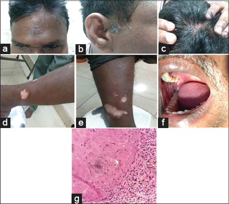
Biopsy from the hyperpigmented plaque showed pseudoepitheliomatous hyperplasia, follicular plugging, civatte bodies, dense mononuclear cell infiltrate in the papillary dermis. Pigment incontinence is also seen (Fig. 1g). Impression was hypertrophic lichen planus. Depigmented patch showed absence of melanocytes and was consistent with vitiligo. Dental opinion was obtained for the oral lesions and a diagnosis of oral erosive lichen planus was made. Patient was prescribed with topical tacrolimus 0.1% and clobetasol propionate for lichen planus and vitiligo lesions respectively. For oral lesions, triamcinolone acetonide (0.1%) for topical application 3 to 4 times a day for 2 weeks half an hour after food was prescribed. Prior to the study, patient gave written consent to the examination and biopsy after having been informed about the procedure.
DISCUSSION
Vitiligo is a common chronic autoimmune disorder affecting the skin and mucous membranes. The depigmentation is due to marked absence of melanocytes and melanin in the epidermis. They can begin at any age but 50% cases develop before age of 20 years. Its exact aetiology is not known but autoimmunity is one of the proposed theories. It has been associated with other autoimmune disorders like diabetes mellitus, thyroid disorder, alopecia areata, pernicious anaemia, myasthenia gravis, addison’s disease and morphoea [2]. In addition, there is presence of melanocyte-specific antibodies detected in vitiligo patients and a higher frequency of organ specific antibodies compared with the general public is observed in such patients [5].
Lichen planus is a chronic inflammatory disorder involving cutaneous and mucosal surfaces, characterized by a T-cell-mediated immune response against epithelial cells, with persistent accumulation of T lymphocytes and epithelial cell damage [6]. The mechanism involved is largely unknown but there is a paucity of immune complexes present in the lesions of lichen planus [7]. There are several variants of lichen planus among which the classical type is the most common followed by the hypertrophic lichen planus and actinic lichen planus. About 30 to 70% of the patients have mucosal involvement [8]. Histologically hypertrophic lichen planus differs from other types by presence of pseudoepitheliomatous hyperplasia of the epidermis and the infiltration being less band like. Direct immunofluorescence shows globular deposits of IgM, and occasionally IgG and IgA, representing apoptotic keratinocytes at the dermoepidermal junction.
Other cases like – actinic lichen planus coexisting with vitiligo; oral lichen planus with vitiligo; becker’s nevus with both vitiligo and segmental lichen planus – has been reported in the literature [9,10].
CONCLUSION
Hypertrophic lichen planus and vitiligo are commonly encountered dermatological entities, but their coexistence is scarcely reported in the literatures. Though the aetiology of both vitiligo and lichen planus is not known with certainty, their coexistence suggests a common autoimmune aetiology.
CONSENT
The examination of the patient was conducted according to the Declaration of Helsinki principles.
REFERENCES
1. Alikhan A, Felsten LM, Daly M, Petronic-Rosic V. Vitiligo: a comprehensive overview: part I. Introduction, epidemiology, quality of life, diagnosis, differential diagnosis, associations, histopathology, etiology, and work-up. J Am Acad Dermatol. 2011;65:473-91.
2. Griffiths C, Barker J, Bleiker T, Chalmers R, Creamer D, editors. Rooks Textbook of Dermatology. John Wiley & Sons; 2016 Feb 29.
3. Petrosian R, Liesionyte K, Petkevicius A, Kucinskiene V, Makstiene J, Valiukeviciene S. Lichen planus verrucosus in an association with vitiligo. Aktuelle Dermatol. 2015;41:425-7.
4. Bhattacharya M, Kaur I, Kumar B. Lichen planus: a clinical and epidemiological study. J Dermatol. 2000;27:576-82.
5. Veitch D, Kravvas G, Hughes S, Bunker C. A rare colocalization of lichen planus and vitiligo. Case Rep Dermatol Med. 2015;2015.
6. Lahlou A, Baybay H, Gallouj S, Mernissi FZ. Childhood vitiligo: Clinical epidemiological profile. Our Dermatol Online. 2017;8:264-7.
7. Oripelaye MM, Olasode OA, Onayemi O, Olanrewaju OF. Familial vitiligo in mother and child; the genetic theory connection. Our Dermatol Online. 2017;8:174-6.
8. Oripelaye MM, Onayemi O, Olasode OA, Olanrewaju FO. Vitiligo on tribal mark: A demostration of Wolf’s isotopic response. Our Dermatol Online. 2017;9:48-50.
9. Mortazavi M, Saad El-Din SA. Bilateral linear lichen planus along the lines of Blaschko: Report of a rare case and brief review. Our Dermatol Online. 2017;8:322-5.
10. Gupta S, Gupta S, Aggarwal K, Jain VK. Becker nevus with vitiligo and lichen planus: Cocktail of dermatoses. North Am J Med Scien. 2010;2:333.
Notes
Source of Support: Nil
Conflict of Interest: None declared.
Comments are closed.