|
Get Citation
|
|
|
El Jouari O, Gallouj S, Douhi Z, Benkirane S, Baybay H, Mernissi FZ. A pregnancy pilomatricoma: an uncommon dermatologic benign neoplasm. Our Dermatol Online. 2018;9(4):425-427. |
|
|
Download citation file:
|
A pregnancy pilomatricoma: an uncommon dermatologic benign neoplasm
Ouiame El Jouari, Salim Gallouj, Zakia Douhi, Selma Benkirane, Hanane Baybay, Fatima Zahra Mernissi
Department of Dermatology and Venereology, University Hospital Hassan II, Fez, Morocco
Corresponding author: Dr. Ouiame EL Jouari, E-mail: eljouariouiame@gmail.com
Submission: 07.01.2018; Acceptance: 31.03.2018
DOI: 10.7241/ourd.20184.17
ABSTRACT
Pilomatricoma is a relatively rare tumour of the skin. It derived from primitive basal cells of epidermis that differentiate into hair matrix cells. It usually arises from the lids and eyebrows. Tumour appear as solitary, firm nodules, exhibiting a normal to pearl white epidermis. A 25-year-old female in her third month of pregnancy, presented with an asymptomatic erythematous nodule 7 weeks. Examination revealed an erythematous nodule, measured approximately 1, 5 cm in diameter firm, with yellowish zones and dotted with telangiectasia. Dermoscopy demonstrated yellowish lobules surrounded by crown-like branching vessels. Patient underwent surgical excision. Microscopic examination revealed a pilomatricoma. After a follow-up of 10 months, the evolution was favorable without recurrence. We report the clinical, dermoscopic, therapeutic and evolutionary characteristics of a case of pilomatricoma in order to help clinicians to better diagnose this entity and decrease the rate of misdiagnosis, especially in pregnant women.
Key words: Pilomatricoma; Periocular; Pregnancy; Dermoscopy
INTRODUCTION
Pilomatricoma is a relatively rare tumour of the skin derived from primitive basal cells of epidermis that differentiate into hair matrix cells. It comprises approximately 1% of all benign skin tumours. Pilomatricoma is an uncommon lesion of the periocular tissues, it usually arises from the lids and eyebrows. Tumour appear as solitary, firm nodules, exhibiting a normal to pearl white epidermis. We describe an interesting case of a pregnancy pilomatricoma arising from the left medial canthus.
CASE REPORT
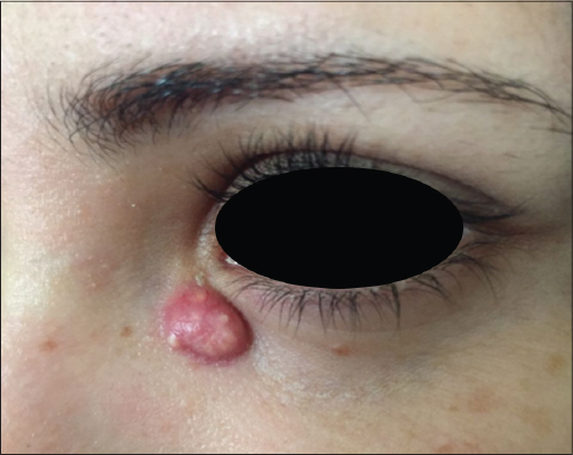
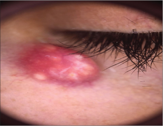
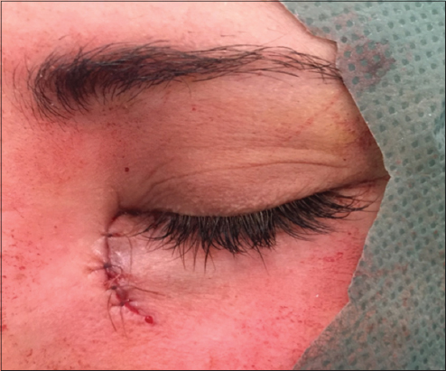
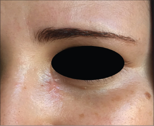
A 25-year-old female in her third month of pregnancy, presented with an asymptomatic erythematous nodule. The patient reported that the lesion started as a small 3-mm papule and grew significantly to a 15 mm lesion in 7 weeks. Examination revealed an erythematous nodule, measured approximately 1, 5 cm in diameter, Limited, with regular contours, firm, with yellowish zones and dotted with telangiectasia (Fig. 1). There was no palpable lymphadenopathy. Dermoscopy demonstrated yellowish lobules on an erythematous background, surrounded by crown-like branching vessels (Fig. 2). Patient underwent surgical excision of the lesion, out under local anaesthesia (Fig. 3). Microscopic examination revealed a regular squamous epithelium which has deep epithelial proliferation mummified cells with a very pale eosinophilic aspect, without having seen any nuclei. Presence of granulomatous reaction giganto-cellular with a foreign body and calcifications (Fig. 4). After a follow-up of 10 months, the evolution was favorable without recurrence (Fig. 5). Prior to the study, patient gave written consent to the examination and biopsy after having been informed about the procedure.

DISCUSSION
Pilomatricoma or calcifying epithelioma of Malherbe is a benign skin tumor. It can occur at any age; congenital forms have been reported. It affects women particularly with a sex ratio of 1.5 [1]. No identified risk factor for the occurrence of pilomatricoma, including pregnancy. The typical clinical aspect of the pilomatricoma is an irregular round or oval asymptomatic subcutaneous nodule of hard or firm consistency. The skin facing the lesion is often bluish. The tumor adheres to the superficial plane, while it is relatively mobile to the deep plane. The most common locations are the head, neck, and upper extremities. There are different clinical forms, perforating, ulcerated, anetodermic with an erythematous skin facing the lesion or pigmented [2].
Dermoscopy help to make the diagnosis, dermoscopic findings of pilomatricoma reported in the literature are yellowish-white structures together with streaks, linear irregular vessels and hairpin like vessels [3,4]. However Confirmation of diagnosis is histological.
Histopathologic examination reveals the tumor to be grossly well circumscribed and firm to gritty in consistency. Microscopic examination shows numerous islands of epithelial cells with characteristic arrangement of basophilic cells in the periphery and shadow cells in the center. As the tumor matures the number of basophilic cells loses their nuclei and becomes shadow cells. Calcification is seen in 75% of the cases. Sheets of intensely eosinophilic keratinous material is seen within necrotic areas, and this may induce a foreign body giant cell reaction [5]. The histopathologic findings of the case described in this reported are similar.
The reference treatment of the pilomatricoma is complete surgical excision, to avoid the risk of recurrence. The prognosis of the pilomatricoma is good [6].
CONCLUSION
Because of the low incidence and variable clinical presentation, pilomatricoma is a tumor not commonly suspected preoperatively. This presentation may help clinicians to better diagnose this entity and decrease the rate of misdiagnosis. Especially in pregnant women.
CONSENT
The examination of the patient was conducted according to the Declaration of Helsinki principles.
REFERENCES
1. Nasreddine FZ, Hali F, Chiheb S. Pilomatricoma: a study of 22 cases. Pan African Med J. 2016;23:254.
2. Ali MJ, Honavar SG. Malherbe’s Calcifying Epithelioma (Pilomatrixoma): An Uncommon Periocular Tumor. Int J Trichology. 2011;3:31–3.
3. Zaballos P, Llambrich A, Puig S, Malvehy J. Dermoscopic findings of pilomatricomas. Dermatology. 2008;217:225–30.
4. Ayhan E, Ertugay OC, Gundogdu R, Three Different Dermoscopic View of Three New Cases with Pilomatrixoma Int J Trichology. 2014;6:21–2.
5. Fernandes BF, Al-Hinai A, Belfort RN, Castiglione E, Arthurs B, Burnier Jr MN, Anetodermic Variant of a Periorbital Pilomatricoma. Ophthal Plast Reconstr Surg. 2008;24:419-21.
6. Marback EF, Cardoso C, Moitinho LM, Marback RL. [Clinicopathologic study of eyelid pilomatrixoma: the experience of the “Hospital Universitario Prof. Edgard Santos.”] Arq Bras Oftalmol. 2007;70:501–3.
Notes
Source of Support: Nil
Conflict of Interest: None declared.
Comments are closed.