|
Get Citation
|
|
|
Mohamed M, Soua Y, Moussa A, Zili J. Linear lichen nitidus: An unusual clinical presentation! Our Dermatol Online. 2017;8(2):218-219. |
|
|
Download citation file:
|
Linear lichen nitidus: An unusual clinical presentation!
Mariem Mohamed1, Yosra Soua1, Adnène Moussa2, Jameleddine Zili1
1Departement of Dermatology, Monastir University Hospital, College of Medicine of Monastir, Monastir, Tunisia; 2Department of Pathology, Fattouma Bourguiba University Hospital, Monastir, Tunisia
Corresponding author: Dr. Mariem Mohamed, E-mail: mariemmohamed79@yahoo.fr
Submission: 01.09.2016; Acceptance: 09.11.2016
DOI: 10.7241/ourd.20172.60
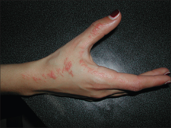
Lichen nitidus (LN) is a rare chronic inflammatory disease of unknown etiology. It predominantly affects children and young adults. Its clinical manifestation typically corresponds to erythematous or flesh-colored, asymptomatic or little pruritic mini papules, often located in the trunk, genitalia, and extremities. The linear arrangement of this dermatosis seems rare. To our knowledge, only 2 cases of linear lichen nitidus have been reported in the literature [1,2]. Our case hence corresponds to the third linear LN case.
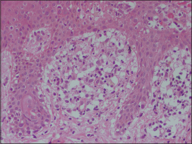
This was a 23-year-old girl with no notable medical history, who consulted for papular discreetly pruritic lesions of the right hand extending to the forearm. The cutaneous examination revealed multiple erythematous millimeters, brilliant mini papules, taking a linear arrangement (Fig. 1). The rest of the integument, the mucous membranes, and the skin appendages were unharmed. The histological examination of a biopsy specimen showed a focally acanthotic epidermis with ortho-hyperkeratosis and a dense inflammatory infiltrate occupying the widened dermal papillae (Fig. 2). The papillary infiltrate was granulomatous comprising lymphocytes, histiocytes and giant cells with the presence of some apoptotic bodies (Fig. 3). In our case, the diagnosis strongly suspected of typical skin lesions was confirmed by the histological study.

REFERENCES
1. Prigent F, Cavelier-Balloy B, Lemarchand-Venencie F, Civatte J. Linear lichen nitidus. Ann Dermatol Venereol. 1989;116:814-5.
2. Petrozzi JW, Shmunes E. Linear lichen nitidus. Cutis. 1970;6:1109-12.
Notes
Source of Support: Nil
Conflict of Interest: None declared.
Comments are closed.