Dermatology eponyms – sign – Lexicon (R): Part 2
Piotr Brzeziński1, Max Carlos Ramírez Soto2,3, Patrice Bourée4, Balachandra S. Ankad5, Shirish Dubey6, Shivanand Gundalli7, Martin G. Baartmans8, Tomasz Wasyłyszyn9, Katarzyna Borowska10, Anca Chiriac11
1Department of Cosmetology, Institute of Biology and Environmental Protection, Pomeranian Academy, Slupsk, Poland; 2Unit Graduate School of Medicine, National University of San Marcos, Lima, Peru; 3Publications Office, National Institute of Health, Peru; 4Parasitic Diseases Unit, Hospital Cochin, 75014 Paris, France; 5Department of Dermatology, S N Medical College, Bagalkot, Karnataka, India; 6Consultant Rheumatologist University Hospital Coventry and Warwickshire NHS Trust, Clifford Bridge Road, Coventry, UK; 7Department of Pathology, S. Nijalingappa Medical College, Bagalkot, Karnataka, India; 8Department of Paediatrics, Maasstadziekenhuis, Rotterdam, Netherlands; 9Department of Dermatology, Military Institute of Medicine in Warsaw, Poland; 10Department of Histology and Embryology with Experimental Cytology Unit, Medical University of Lublin, Poland; 11Department of Dermatology, Nicolina Medical Center, Iasi, Romania
Corresponding author: Piotr Brzezinski, MD, PhD., E-mail: brzezoo77@yahoo.com
Submission: 22.08.2016; Acceptance: 01.12.2016
DOI: 10.7241/ourd.20171.35
ABSTRACT
Eponyms are used almost daily in the clinical practice of dermatology. And yet, information about the person behind the eponyms is difficult to find. Indeed, who is? What is this person’s nationality? Is this person alive or dead? How can one find the paper in which this person first described the disease? Eponyms are used to describe not only disease, but also clinical signs, surgical procedures, staining techniques, pharmacological formulations, and even pieces of equipment. In this article we present the symptoms starting with (R) and other. The symptoms and their synonyms, and those who have described this symptom or phenomenon.
Key words: Eponyms; Skin diseases; Sign; Phenomenon
Rinman’s Sign
The appearance of cord like radiations proceeding from the nipple. An early sign of pregnancy.
Ringworm Sign
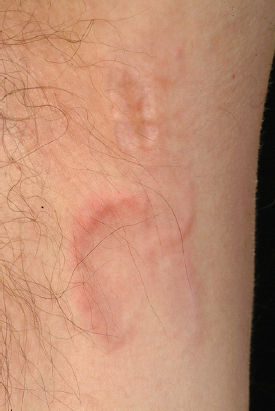
Tinea corporis (“ringworm”): Note the outer red border and clear center (Fig. 1). T. rubrum is the most common cause [1].
Ritter’s Sign
Dermatitis exfoliativa neonatorum (Fig. 2), with diffuse and universal scaling, which may be branny or in laminae like pitiriasis rubra [2,3].
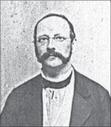
Gottfried Johann Nepomucenus Ritter von Rittershain
German physician and pediatrician, 1820-1883 (Fig. 3). He studied at Lviv University and Charles University. The title of his thesis was “De epilepsia”. He worked at the clinic in Prague and was interested in episode paediatrics. In 1864 he was appointed professor of pediatrics in Prague. He was founder of the Prague medical weekly. In 1880 he had to give up his job because of his epilepsy. He died on August 20, 1883 of a stroke [4]. On Ritter is the Ritter disease, so the staphylococcal scalded skin syndrome.
Rivolta’s Sign
Actinomycosis (Fig. 4). The term “Actinomycosis” was derived from the Greek terms aktino, which refers to the radiating appearance of a sulfur granule, and mykos, which labels the condition a mycotic disease [5].
Sebastiano Rivolta
Italian veterinarian and bacteriologist, (1832-1893) (Fig. 5). He was persuaded by his father to join the Royal School of Veterinary Medicine of Turin in 1852, winning a scholarship and graduating with honors four years later.
He immediately set out for his intensive research in parasitology, bacteriology, pathology and clinical anatomy, which led to the discovery and observation of the agents responsible for numerous veterinary diseases. Among them we can mention the Discomyces bovis (1868); Cryptococcus farciminosus (1873); Avitellina centripunctata (1874); Discomyces equestris (1884); Opisthorchis felineus (1884).
With his studies contributed to a greater understanding of the role of many pests germs from income and not: Demodex folliculorum; Moniezia expansa; Taenia echinococcus; Bacillus anthracis. First he observed the cellular inclusions characteristic of diftero-avian pox, laying the foundation for the study of this disease caused by a virus, in a period when the virology was yet to be born. To him we owe the discovery of some special cells in the retina of the horse, so called “cells of the Revolt”.
The experience allowed him to teach six different materials at the Turin University, before moving to the University of Pisa. He became a full professor of General Pathology and Veterinary Pathological Anatomy, practiced teaching in the Tuscan city for 22 years, until his death occurred as a result of complications of his precarious state of health, due to a severe chronic disease that accompanied him from his youth. It is still buried in the monumental cemetery of Alexandria [6].
Robinson’s Sign
Hidrocystoma (Fig. 6a and b) [7].
Andrew R. Robinson
American dermatologist, 1845.
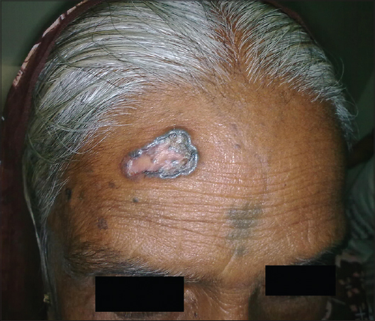
Rodent Ulcer Sign
Malignant ulcer situated above a line which joints the angle of the mouth and the tragus of the ear. Called a rodent ulcer because the wound looks like a rat has gnawed at the tissue and bone (Fig. 7) [8].
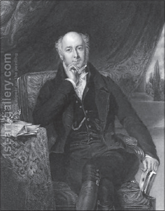
Sir Charles Mansfield Clarke
(1st Baronet) English physician, 1782-1857 (Fig. 8). After leaving St Paul’s School, he received his medical training at St George’s Hospital and the Hunterian School of Medicine. He spent two years as assistant surgeon in the Hertfordshire Militia. He left the army and, specialised in midwifery and in women’s and children’s diseases. Between the years 1804 and 1821, he delivered regular courses of lectures on these subjects. His reputation as a practitioner during these years reached great heights and numerous honours were bestowed on him, including the Fellowship of the Royal Society in 1825, the appointment of Physician to Queen Adelaide in 1830, a baronetcy in 1831, and honorary degrees at Cambridge and Oxford in 1842 and 1845. He was president, and an enthusiastic supporter, of the Society for the Relief of the Widows and Orphans of Medical Men [9].
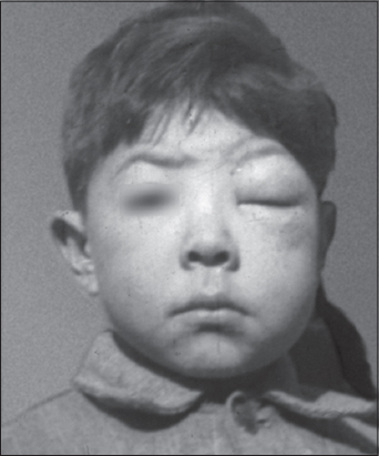
Romaňa’s Sign
Romaňa’s sign is the first symptom of american trypanosomiasis (or Chagas’ disease) (Fig. 9). When the route of inoculation of parasites (Trypanosoma cruzi) is the ocular mucosa, edema of the eyelids and conjunctivitis may occur. This unilateral periorbital edema which does not pit on pressure and with a dry skin is thought to be pathognomonic for early Chagas’disease. Chagas’disease is a zoonosis, caused by Trypanosoma cruzi, which was discovered by Carlos Chagas in Brazil in1909. About 18 million persons are infected in south America, mostly in Brazil and Argentina [9,10].
Cecilio Felix Romaňa
Argentinean physician (1899-1997). He was an Argentinean researcher dedicated to tropical diseases firstly in the area of Santa Fe then in Oswaldo Cruz Institute (Rio da Janeiro) with S. Mazza. Romaňa became famous when he described this symptom in 1935. But, the director of the Institue, S. Mazza, never accepted neither the specificity of this sign nor its popular name as Romaňa’s sign [10].
Rhomboid Sign
Central papillary atrophy of the tongue, associated with the presence of Candida albicans [11,12].
Rope Sign
It refers to the thick indurated inflammatory cord like structure that extends from the lateral trunk to the axillae and said to be a classical finding of interstitial granulomatous dermatitis (Ackerman’s syndrome) with arthritis (Fig. 10) [13].
Rose Handler’s Sign
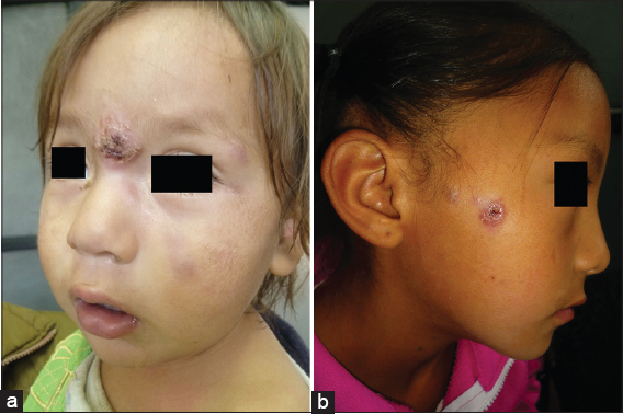
Ulcerative skin lesions with nodular lymphangitis, due to exposure to the zoonotic fungal Sporothrix schenckii, found on peat moss, horses, laboratory animals, other mammals, and birds (Fig. 11a and b) [14].
Rose Spots Sign
Rose spots on the abdomen with typhoid fever [15].
Rosenbach’ Sign
- Absence of the abdominal skin reflex in inflammatory disease of the intestines.
- Absence of the abdominal skin reflex in pinching the skin of the abdomen on the paralyzed side in hemiplegia.
- A fine rapid tremor of the closed eyelids in Graves’ disease.
- Inability to close the eyes immediately on command; seen in neurasthenia.
Ottomar Ernst Felix Rosenbach
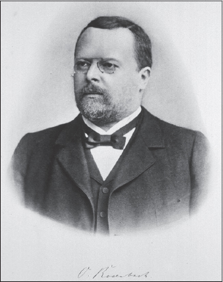
German physician, 1851-1907 (Fig. 12). He received his education at the universities of Berlin and Breslau (M.D. 1874). His studies were interrupted by the Franco-Prussian war, in which he took an active part as a volunteer. From 1874 to 1877 he was assistant to Wilhelm Olivier Leube and Carl Wilhelm Hermann Nothnagel at the medical hospital and dispensary of the University of Jena; in 1878 he was appointed assistant at the Allerheiligen-Hospital at Breslau, and became privatdozent at the university of that city; in 1887 he became chief of the medical department of the hospital, which position he resigned in 1893; and in 1888 he was appointed assistant professor. In 1896 he resigned his professorship and removed to Berlin, where he practised until his death.
He discovered unusual eye tremors when the eyelids are closed in patients with Graves disease. He also described a clinical sign for aortic regurgitation (involving systolic pulsations of the liver) [16].
Ross River Rash Sign (Australia, South Pacific Islands)
Purpura on the lower extremities with fever and polyarthritis. Caused by the mosquito-borne zoonotic Ross River alphavirus [17].
Rössle’s Sign
Plantar hypersensitivity. A sign in thrombosis [18].
Robert Rössle
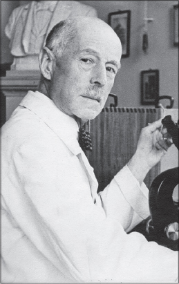
German pathologist, 1876-1956 (Fig. 13). In 1900 he received his medical doctorate from Munich, and went to work at the Pathological Institute of the University of Kiel. From 1911 to 1921, he was a professor of general pathology and pathological anatomy at the University of Jena, and from 1922 until 1929 he held a similar position in Basel. In 1929 he succeeded Otto Lubarsch in the department of pathology at the Charité in Berlin, where he remained until 1948.
Rössle performed pathological investigations in several facets of medicine, including liver disease, allergies, inflammation, cellular pathology and geriatrics. He described aspects associated with a form of secondary biliary cirrhosis that was once referred to as “Hanot–Rössle syndrome” (named in conjunction with French physician Victor Charles Hanot).
Rössle published over 300 medical papers, and was editor of 39 volumes of Virchow’s Archiv für Pathologische Anatomie und Physiologie und für Klinische Medizin (now: Virchows Archiv). Today, the Robert-Rössle-Hospital and Tumor Institute is named in his honor. It is located at the Max-Delbrück-Center of Molecular Medicine at the Humboldt University of Berlin [18].
Rostan’s Sign
Gangrene of the penis associated with small pox.
Léon Louis Rostan
French physician, 1790-1866 (Fig.14). He studied medicine in Marseilles and Paris. He was a disciple of Philippe Pinel, and for much of his professional career was associated with the Pitié-Salpêtrière Hospital in Paris.
In 1819 Rostan was the author of Recherches sur le ramollissement du cerveau (Researches on cerebral softening), in which he provided the first accurate description of spontaneous cerebral softening. He documented that the disorder was a specific anatomo-clinic entity that was different from encephalitis and apoplexy. His findings were harshly criticised by followers of Broussais’ teachings on physiological medicine, who claimed that brain softenings were the result of an inflammation process, and therefore should be depicted as encephalitis.
He also did extensive research of animal magnetism and somnambulism, and wrote a treatise on charlatanism for his graduate thesis. Rostan performed early studies on the classification of body types, using descriptive terms such as respiratory-cerebral, muscular and digestive in his analysis.
In 1845, he was elected a foreign member of the Royal Swedish Academy of Sciences [19].
Rothschild’s Sign
Loss of hair from lateral third of eyebrows in hyperthyroidism [20].
Rumpel-Leede Sign
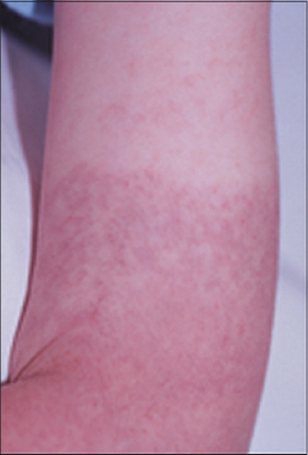
The appearance of minute subcutaneous haemorrhages below a bandage applied on the upper arm for ten minutes (Fig. 15). A sign of haemorrhages diathesis, capillary fragility, and scarlet fever [21]. Also known as Hechtsign.
Carl August Theodor Rumpel
German surgeon, 1862-1923. He was remembered for describing the Rumpel-Leede sign.
He received his doctorate in 1887 in Marburg and worked at the Hamburg-Eppendorf Hospital and took part in the fight against the cholera epidemic of 1892. He oversaw the building of the Barmbecker Krankenhaus in Hamburg, of which he became director in 1913.
With internist Alfred Kast, he was co-author of a patho-anatomical atlas titled: “Pathologisch-anatomische Tafeln nach frischen Präparaten mit erläuterndem anatomisch-klinischem” [22].
Carle Stockbridge Leede
German-American physician, 1882-1964 (Fig. 16). Physician at the Children’s Hospital in Seattle.
Russell’s Sign
Lanugo, dry skin, and hand calluses, associated with purgino and bulimia [23].
Gerald Francis Morris Russell
British psychiatrist, born in 1928. In 1979 he published the first description of bulimia nervosa, and Russell’s sign has been named after him. Russell was schooled at George Watson’s College, Edinburgh, and he qualified as a medical doctor with BM BCh from the University of Edinburgh in 1950.
From 1971 to 1979 Russell was a professor and consultant psychiatrist at the Royal Free Hospital, London, and from 1979 to 1993 he was a professor at the Institute of Psychiatry at the Maudsley Hospital, London, where he set up an eating disorder unit, which has been named after him. From 1993 he has worked at Priory Hosp Hayes Grove, Bromley, Kent.
Russian Horse Sign
Glanders, a zoonotic bacterium Burkholderia mallei, that causes skin and mucous membrane lesions, as well pneumonia. The bacteria has been used as a biological warfare weapon in Word Wars I and II [24].
ACKNOWLEDGEMENTS
Giuseppina Piccolo from Biblioteca di medicina veterinaria Via Celoria, 10 20133 Milano, Italy, for the information about Sebastiano Rivolta.
REFERENCES
1. Chiriac A, Brzezinski P, Chiriac AE, Pinteala T, Mares M, Moldovan C, Why Is Tinea an Annular Lesion With Centrifugal Growth?Int J Surg Pathol 2015; 23: 652-3.28.
2. Oishi T, Hanami Y, Kato Y, Otsuka M, Yamamoto T, Staphylococcal scalded skin syndrome mimicking toxic epidermal necrolysis in a healthy adultOur Dermatol Online 2013; 4: 347-8.
3. Baartmans MG, Dokter J, den Hollander JC, Kroon AA, Oranje AP, Use of skin substitute dressings in the treatment of staphylococcal scalded skin syndrome in neonates and young infantsNeonatology 2011; 100: 9-13.
4. Oehme J, [Pioneers in pediatrics. Gottfried Ritter von Rittershain (1820-1883)]Kinderkrankenschwester 1990; 9: 334-
5. Wasyłyszyn T, Borowska B, Cutaneous actinomycosis. A case reportOur Dermatol Online 2016; 7: 450-1.
6. Theodorides J, Sebastiano Rivolta (1832-1893) parasitologiste et microbiologisteHist Scien Med 1980; 14: 227-32.
7. Ankad BS, Domble V, Sujana L, Dermoscopy of apocrine hydrocystoma:A first case reportOur Dermatol Online 2015; 6: 334-6.
8. Gundalli S, Kolekar R, Kolekar A, Nandurkar V, Pai K, Nandurkar S, Study of basal cel carcinoma and its histopathological variantsOur Dermatol Online 2015; 6: 399-403.
9. Brzezinski P, Passarini B, Nogueira A, Dermatology eponyms – phenomen/sign – dictionary (C)N Dermatol Online 2011; 2: 81-100.
10. Bourée P, Romana’s signOur Dermatol Online 2015; 6: 248-
11. Al Aboud K, Al Aboud A, Eponyms in the dermatology literature linked to Latin AmericaOur Dermatol Online 2013; 4: Suppl.1405-8.
12. Farman AG, van Wyk CW, Staz J, Hugo M, Dreyer WP, Central papillary atrophy of the tongueOral Surg Oral Med Oral Pathol 1977; 43: 48-58.
13. Dubey S, Merry P, Clinical vignette. Interstitial granulomatous dermatitis (Ackerman’s syndrome) in SLE presenting with the ’rope sign’Rheumatology (Oxford) 2007; 46: 80-
14. Ramírez Soto MC, Facial Sporotrichosis in children from endemic area in PeruOur Dermatol Online 2013; 4: 237-40.
15. Raveendran KM, Viswanathan S, Typhoid Fever, Below the BeltJ Clin Diagn Res 2016; 10: OD12-3.
16. Temmen H, [Ottomar Rosenbach 1851-1907) and his contribution to psychotherapy]Acta Psychother Psychosom 1964; 12: 10-20.
17. Reusken C, Cleton N, Medonça Melo M, Visser C, GeurtsvanKessel C, Bloembergen P, Ross River virus disease in two Dutch travellers returning from Australia, February to April 2015Euro Surveill 2015; 20: 21200-
18. Schlag PM, [Hans Gummel on the 30th anniversary of his death. 1954-1973 chief surgeon and director of the Robert-Rössle Clinic]Chirurg 2004; 75: 300-1.
19. Poirier J, Chretien F, Léon Rostan (1790-1866)J Neurol 2000; 247: 659-60.
20. Jordan DR, Ahuja N, Khouri L, Eyelash loss associated with hyperthyroidismOphthal Plast Reconstr Surg 2002; 18: 219-22.
21. Brzezinski P, Pessoa L, Galvão V, Barja Lopez JM, Adaskevich UP, Niamba PA, Dermatology Eponyms – sign – Lexicon (H)Our Dermatol Online 2013; 4: 130-43.
22. Pieper C, Die Sozialstruktur der Chefärzte des Allgemeinen Krankenhauses Hamburg-Barmbek 1913 bis 1945LIT Verlag Münster 2003;
23. Young J, Henderson MC, Thompson GR, 3rdAnorexia nervosa:Russell’s sign with concurrent tetanyJ Gen Intern Med 2011; 26: 668-
24. Jones BV, Glanders and historyVet Rec 2016; 178: 664-
Notes
Source of Support: Nil
Conflict of Interest: None declared.






Comments are closed.