|
Get Citation
|
|
|
Gupta M. Angiolymphoid hyperplasia with eosinophilia. Our Dermatol Online. 2017;8(1):94-95. |
|
|
Download citation file:
|
Angiolymphoid hyperplasia with eosinophilia
Mrinal Gupta
Consultant Dermatologist, Treatwell Skin Centre, Jammu, India
Corresponding author: Dr Mrinal Gupta, E-mail: drmrinalgupta@yahoo.com
Submission: 10.05.2016; Acceptance: 16.08.2016
DOI: 10.7241/ourd.20171.24
Angiolymphoid hyperplasia with eosinophilia (ALHE), also called as atypical or pseudo pyogenic granuloma, is characterized by solitary or multiple red to brown papules or nodules seen commonly in women between 20 and 40 years of age. The etiology of ALHE remains unknown, because it is not clear if it is primarily a vascular neoplasm, a lymphoproliferative process or a heterogeneous group of entities [1]. Trauma, infections and hyperestrogenic conditions (pregnancy or oral contraceptive agents) are considered to be the likely causes. ALHE usually appears in head and neck region, frequently in the auricular area and usually measures about 2-3 cm in size [2]. ALHE must be histologically and clinically differentiated from Kimura disease, which is a chronic inflammatory condition characterized by large subcutaneous nodules in the head and neck region [3].
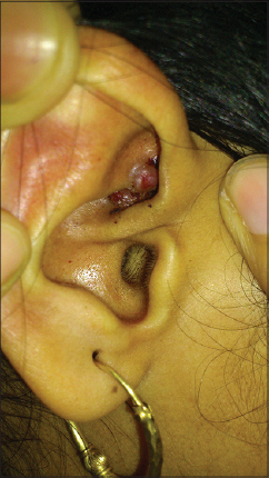
A 15 year old male presented to us with history of asymptomatic, gradually progressive nodular lesions over the meatus of the right ear. The lesions started at the age of 11 years and were a single nodule to begin with, but over the time, they had increased in size and number to their present size, but had remained asymptomatic throughout. The patient had consulted some physicians and had applied numerous medications without any relief. On examination, there were multiple red, non-pulsatile smooth surfaced papules, over the pre auricular region extending on to the crus of the helix, and into the meatus of the right ear (Fig. 1). There was no lymphadenopathy. Other systems were normal. Differential count showed 16% eosinophils and her IgE level was normal.
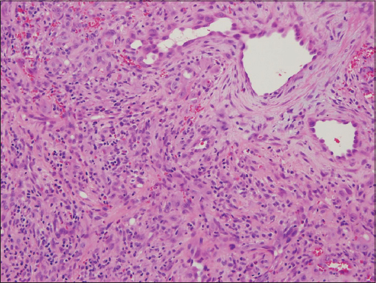
An excisional biopsy of the lesions was done which revealed proliferation of small vessels lined by plump endothelial cells, surrounded by inflammatory cells including lymphocytes, mastocytes and eosinophils which confirmed the diagnosis of ALHE (Fig. 2).
REFERENCES
1. Karabudak O, Taskapan O, Bozdogan O, and Dogan B, Angiolymphoid hyperplasia with eosinophilia:Atypical appearance in an older patientIndian J Dermatol 2008; 53: 144-5.
2. Zarrin-Khameh N, Spoden JE, Tran RM, Angiolymphoid hyperplasia with eosinophilia associated with pregnancy:A case report and review of the literatureArch Pathol Lab Med 2005; 129: 1168-71.
3. Zaraa I, Mlika M, Chouk S, Chelly I, Mokni M, Zitouna M, Angiolymphoid hyperplasia with eosinophilia:A study of 7 casesDermatol Online J 2011; 17: 1-
Notes
Source of Support: Nil
Conflict of Interest: None declared.
Comments are closed.