|
Get Citation
|
|
|
Bouhaja L, Jones M, Rammeh S. Folliculosebaceous cystic hamartoma. Our Dermatol Online. 2016;7(4):477-478. |
|
|
Download citation file:
|
Folliculosebaceous cystic hamartoma
Leila Bouhaja1, Meriam Jones2, Soumaya Rammeh1
1Department of Pathology, Charles Nicolle Hospital, Tunis, Tunisia, 2Department of Dermatology, Charles Nicolle Hospital, Tunis, Tunisia
Sir,
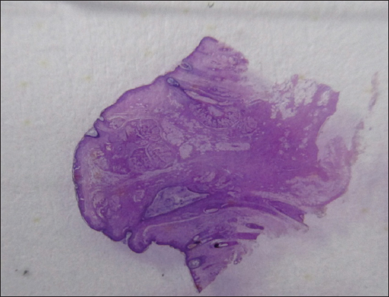
We report a 34 year-old man, with no past medical history, who presented with a 2-year history of a nodule on the nose. At physical examination the lesion was firm, pedunculated, and flesh-colored with a smooth surface. Clinically, the diagnosis of adnexal tumor was suggested and a surgical resection of the lesion was performed. Grossly, the tumor was firm pedunculated measuring 6 mm in size, raising the epidermis (Fig. 1). Histological examination showed a well-circumscribed lesion of the dermis composed of numerous sebaceous follicles structures. Some were dilated forming cysts and connected directly to the epidermis (Fig. 2). These epithelial structures were surrounded by a fibrohyalin stroma combined with a fibrous component containing adipocytes (Fig. 3). These features were reminiscent with the diagnosis of folliculosebaceous cystic hamartoma. At 10 months of follow-up, the patient was asymptomatic and had no recurrence.


The patient’s informed consent was obtained.
Prior to the study, patient gave written consent to the examination and biopsy after having been informed about the procedure.
Folliculosebaceous cystic hamartoma (FSCH) is a rare cutaneous hamartoma composed of dilated folliculosebaceous units associated with mesenchymal elements [1]. This tumor may occur in all age groups, and females are affected more often than males [2]. Clinically, it manifests as a solitary skin-colored sessile or pedunculated nodule that is most commonly located on the face and scalp, with approximately 30% occurring in the nasal or paranasal regions. The lesion is usually less than 3 cm in size [3,4]. The main differential diagnoses are nevus lipomatosus superficialis or “sebaceous” trichofolliculoma which may include sebaceous glands but no central cyst [5]. FSCH can be readily identified by the presence of adipocytes and a fibrous stroma. The treatment consists of total excision of the tumor.
Consent
The examination of the patient was conducted according to the Declaration of Helsinki principles. Written informed consent was obtained from the patient for publication of this article.
REFERENCES
1. Osipov VO, Vincent P, Packer AM, Oliver GF, Folliculosebaceous cystic hamartoma of the ear and periauricular skinAustral J Dermatol 2012; 53: e8-e9.
2. Ansai S, Kimura T, Kawana S, A clinicopathologic study of folliculosebaceous cystic hamartomaAm J Dermatopathol 2010; 32: 815-20.
3. Watanabe-Okada E, Kurihara Y, Miyakawa S, Tanaka M, Dermatoscopy of folliculosebaceous cystic hamartomaDermatol Pract Concept 2014; 4: 47-9.
4. Nguyen CM, Skupsky H, Cassarino D, Folliculosebaceous Cystic Hamartoma With Spindle Cell Lipoma-Like Stromal FeaturesAm J Dermatopathol 2015; 37: e140-2.
5. Merklen-Djafri C, Batard M.-L, Guillaume J-C, Kleinclauss I, Cribier B, Folliculosebaceous cystic hamartoma: Anatomo-clinical studyAnn Dermatol Venereol 2012; 139: 23-30.
Comments are closed.