|
Get Citation
|
|
|
Choccalingam C. Hibernoma of cervical region. Our Dermatol Online. 2016;7(4):475-476. |
|
|
Download citation file:
|
Hibernoma of cervical region
Chidambharam Choccalingam,
Department of Pathology, Chettinad Medical College, Kelambakkam, Tamil Nadu, India
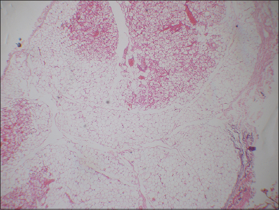
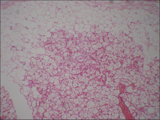
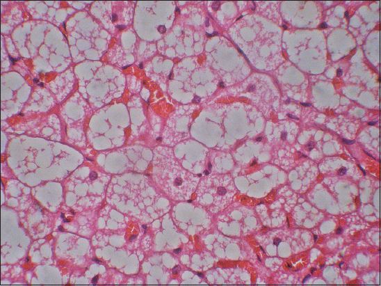
A 42 year old male presented with swelling in the right posterior cervical region for a period of 2 months. Palpation revealed a soft, non tender, mobile swelling measuring 2 x 1 cms. An elective excision biopsy was performed. Gross examination showed a yellow to pale brown soft mass. On microscopy, a well circumscribed tumour composed of two populations of cells(uni-vacuolated white adipocytes and multi-vacuolated brown adipocytes) was seen (Fig. 1). Gradual transition between the two populations was seen (Fig. 2). The white adipocyte cells with clear cytoplasm and peripheral nucleus were seen in the periphery of tumour. The brown adipocyte cells with granular eosinophilic multi-vacuolated cytoplasm and central round vesicular nucleus were seen in the central areas of tumour (Fig. 3). No atypia or necrosis or mitosis was seen. With the morphological features a diagnosis of hibernoma- benign tumour of brown fat was made.

Hibernoma is a benign tumour of brown fat that is also known as fetal lipoma, lipoma of embryonic fat and usually is seen in interscapular, neck, axilla, mediastinal and retro-peritoneal regions of adults where vestiges of brown foetal fat persists beyond foetal life [1,2]. Brown adipocyte characterized by small lipid droplets, high mitochondrial content is seen mostly in newborns and in hibernating mammals and is postulated to play a role in thermoregulation [3]. The high iron content within the mitochondria and the rich blood supply imparts the brown colour to the hibernoma [1,3]. Microscopically, presence of brown adipocyte cells is common to all the four variants namely typical(lobular pattern with uni-vacuolar and multi-vacuolar adipocytes), myxoid (prominent myxoid change), lipoma like (predominantly and spindle cell type. Surgical resection of tumour is curative with no recurrence or transition to malignancy [1].
REFERENCES
1. Weiss SW, Goldblum JR, Enzinger and Weiss’s Soft tissue Tumours 2008; 5 ed. Mosby, Inc;
2. Furlong M, Fanburg-Smith J, Miettinen M, The morphologic spectrum of hibernoma: a clinicopathologic study of 170 casesAm J Surg Pathol 2001; 25: 809-14.
3. Chen C-L, Chen W-C, Chiang J-H, Ho C-F, Interscapular hibernoma-Case report and literature reviewKaohsiung J Med Sci 2011; 27: 348-52.
Notes
Source of Support: Nil
Conflict of Interest: None declared.
Comments are closed.