Eyelid nonbullous impetigo
Rafael Corredor-Osorio, Martha Corredor-Osorio, Alcira Corredor-Osorio
Corredor Ocular Center Specialized Ophthalmology. Av. Bolívar, CC Las Acacias, local 31, Valera (Trujillo), Venezuela
ABSTRACT
We report an unusual case of nonbullous impetigo of the eyelid. A 21-year-old woman had a painless eruptions right eyelid and around nostril. Clinical examination revealed eyelid nonbullous impetigo. The patient was otherwise healthy. Any minor trauma or skin problems there were not founded. Staphylococcus aureus was isolated from the lesions. Complete resolution was achieved with oral cephalexin an ointment tobramycin. Impetigo nonbullous occurs commonly on the face of children but it might be developed in healthy adults even those was not necessary evidenced previous trauma or dermal infiltrate with pus or any other active cutaneous injury as this case is demonstrated. The treatment of impetigo around the eye included a careful examination of the segment anterior and the application de topical measures with oral and topical antibiotics. The case is being reported in view of its rarity and unusual location.
Key words: Impetigo; Staphylococcus aureus; Eyelid; Streptococcus pyogenes
INTRODUCTION
Impetigo is a highly contagious bacterial skin infection affecting children [1–4] worldwide that is caused by Gram-positive bacteria Staphylococcus aureus [5–7] Streptococcus pyogenes [2,4] or both [2,8–10].
The face, especially the perioral region, is one of the most frequently involved areas [2]. The infection usually begins with an outbreak of vesicular lesions on the arms and legs and on occasion they are seen around the nose, mouth and scalp [11]. Here we report an unusual case of nonbullous impetigo in a woman who presented crusting, vesiculobullous skin lesion in the right upper eyelid.
CASE REPORT
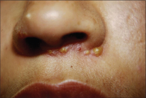
A 21-year old woman present with a lesion in right eyelid. It began a week earlier as an erythematous macule that soon becomes vesicular that rupture, ooze, and lead to the layer of crusting. There was no history of ocular trauma or history of any surgery performed in the past. No family or personal history of skin problems. The clinical appearance was scattered, painless, slightly pruritic reddened sores with honey–yellow exudates crusting on them, localized in upper eyelid (Fig. 1). Satellite lesion was observed around the nose (Fig. 2). There was region auricular lymphadenopathy. The eyelid culture was positive for S. aureus. The remainder of the ocular and general physical examination was normal. Treatment was initiated with oral cephalexin mono hydrate 500mg per each 12 hours, ointment tobramycin four times daily. The patient responded well to the antibiotic, and the denuded area showed slow epithelialization and complete healing of the skin within 3 weeks.

DISCUSSION
Impetigo is a common localized crusting, vesiculobullous skin lesion resulting from superficial infection by bacteria. The bacteria usually infect skin that has been damaged by scratching an insect bite or picking a scab. The lesions may cause mild soreness an itching, but are typically painless. Impetigo was described in 1864 by Tilbury–Fox. The isolated micro-organism usually identified in cultures of lesions is a S. aureus [5–7] although S. pyogenes infection is also common [2,4]. Many lesions represent a mixture of the two infections [2,8–10].
Susceptibility to these infections depends on host immune factors, as well as virulence of the organism. It is most likely to occur under conditions of crowding, poor hygiene, and hot, humid climate, and it can spread rapidly between members of a household, school. Impetigo can occur as a primary infection or secondary to pre-existing skin conditions, such as eczema or scabies [3]. Impetigo can be divided on clinical and bacteriological grounds into basic forms: Impetigo non-bullous (contagiosa)and bullous based on the presence or lack large blisters, called bullae [2,4,9,10]. Both forms involve only the most superficial layers of the skin [10]. Impetigo nonbullous is the most common form and is likely to be caused by a mixed staphylococcal and streptococcal infection [2,9,10] depending on geographical variations, with the streptococcal form being more prevalent in warmer and humid climates[8], however, S. aureus in the main cause. The early lesion of impetigo non-bullous begins as a small, 2-4 mm, erythematous macule which soon becomes vesicular or pustular [12].
The vesicle is a very thin-walled hardly noticeable, as it soon ruptures, leaving an exudate [4,12]. The purulent exudates dries and form the classic thick yellow-brown crust (meliceric)[3,4] with a honey color [2,8,9]. The crusts lesion, may show no residual or active vesicles and measures 1-3 cm, in greatest diameter. It may be mildly pruritic. Satellite lesions occur in the vicinity due to autoinoculation [2]. When removed, the crust quickly reappears [4]. Removal of the crust results in the reaccumulation of fresh exudates. The crusts eventually dry separate and disappear, leaving an area of erythema that heals without scarring.
The lesions are painless and usually localized, often occur in the face around the mouth and nose. The trunk and limbs can also be affected. Systemic signs are usually not present however with extensive impetigo, fever and regional lymphadenopathy may occur [2].
Bullous impetigo is almost always caused by S. aureus [2,4,5,9] is characterized by erythematous macule that progress to bullae, large thin-walled blister that contain clear or cloudy yellow fluid [2,4], and measure less that 5cm in diameter [10]. Bullae are caused by staphylococcal infections. Staphylococcally produced epidermolytic toxin has been recovered from the blister fluid in some cases [9,13]. These blisters easily rupture and leave behind a moist area of eroded skin surrounded by a thin ring of the remaining blistered skin [2]. The lesion dries and crust over, creating a light brown appearance that resembles “varnish” which may be white or grey in color [11]. These lesions are discrete, with little redness or inflammation surrounding them [2]. Its usual distribution involves the face, buttocks, truck and perineum [3]. The condition might be associated with fever, diarrhea and weakness [10]. Although the nonbullous impetigo is a dermatological disease may be associated with conjunctivitis [14]. Most cases resolve without sequelae in 2 to 3 weeks. Complications of impetigo include cellulitis, lymphangitis, supurative lymphadenitis, nephritis and sepsis. The differential diagnosis of nonbullous impetigo should include herpes simplex viral infections, herpes zoster, candidiasis, atopic contact dermatitis, seborrheic dermatitis, insect bites, varicella, scabies and burns thermal [4,10]. The aim of treatment is to clear the eruption and prevent the spread of one infection to other. Nonbullous impetigo can be treated with either oral or topical antibiotics. At the initiation of treatment the crusty scabs should be removed after softening with wet compresses. The lesion should be washed with an antibacterial soap [4] or povidine-iodone shampoo two or three times daily [3]. Initial cultures are usually not necessary, since health care professional commonly prescribe erythromycin [2–4,8], amoxicillin/clavulanate [2,3], cephalexin [2,3,10], vancomycin [1,2], azithromycin, oxacycline [4], dicloxacillin [14] and clindamycin [2]. The course of treatment is 7 days.
For mild infections, a topical antibiotic efficacious against gram-positive bacteria, especially S. aureus and S. pyogenes is the preferred first-line therapy. Topical treatment may be selected if area of impetigo is localized. An antibiotic ointment or cream should be then applied, such as mupirocin 2% ointment [1,4,8], neomycin, bacitracin ointment [2,3], chloramphenicol ointment [14] fusidic acid cream 2% [1,4,6], gentamycin ointment [3], retapamulin 1% ointment [1,4,6], NVC-422 topical gel [1], sulphadiazine cream [11]. This approach is sufficient to clear mild to moderate cases. It is of interest that the patient developed localized crusting, vesiculobullous skin lesion, clinically similar a nonbullous impetigo characterized for several adjacent clusters of small clear vesicles and whitish pustules which are partly confluent and is yellow-brown break open early to release a clear, oozing secretion which quickly becomes crusted on the surface. The crusted lesion is honey color. This lesion is localized in the right upper eyelid, although the upper lip, labial commisure, suborbital skin are affected more often. For widespread, localized infections, such as this patient, require oral and topical antimicrobials to be sure that infection has not worsened and developed into skin infection called cellulitis.
In summary, impetigo non bullous is a common, vesiculopustular, crusting skin lesion resulting from superficial infection by bacteria. Typically affects children but it can affect age group. Intact, healthy skin seems largely immune to the disease, as most lesions occur in areas of dermatitis or previous trauma and, is usually transmitted direct skin contact or indirect contact with the liquid ooze of an lesion active. When impetigo non bullous occurs around of the eye especially in the eyelids, a careful examination of the anterior segment of the eye is necessary and, if are signs of increased redness, or pain in the skin surrounding of the lesion, oral and topical antibiotics should be used for avoid potential complications. Ophthalmologists, dermatologists, and physicians should be familiar with this entity of impetigo and consider it in the differential diagnosis of the spectrum of the vesicobullous disorders in the eyelid.
Consent
The examination of the patient was conducted according to the Declaration of Helsinki principles written informed consent was obtained from the patient for publication of this article and any accompanying image.
REFERENCES
1. Lovino SM, Krantz KD, Blanco DM, Fernández JA, Ocampo N, Najafi A, NVC-422 topical gel for the treatment of impetigoInt J Exp Pathol 2011; 4: 587-95.
2. Motswaledi MH, Impetigo in children: a clinical guide and treatment optionsS Afr FAM Pract 2011; 53: 44-6.
3. George A, Rubin G, A systematic review and meta-analysis of the treatment for impetigoBr J Gen Pract 2003; 53: 480-7.
4. Empinotti JC, Uyeda H, Ruaro RT, Galhardo AP, Bonatto DC, PyodermitisA Bras Dermatol 2012; 87: 277-84.
5. Cho JS, Zussman J, Donegan NP, Ramos RI, Garcia NC, Uslan DZ, Noninvasive in vivo imaging to evaluate immune responses and antimicrobial therapy against staphylococcus aureus and USA300 MRSA skin infectionsJ Invest Dermatol 2011; 131: 907-15.
6. Alsterholm M, Flystrom I, Bergbrant IM, Faergeman J, Fusidic acid-resistant staphylococcus aureus in impetigo contagiosa and secondarily infect atopic dermatitisActa Derm Venereol 2010; 90: 52-7.
7. Shi D, Higuchi W, Takano T, Saito K, Ozaki K, Takano M, Bullous impetigo in children infected with methicillin-resistant staphylococcus aureus alone or in combination methicillin-susceptible S. aureus: analysis of genetic characteristics, including assessment of exfoliative toxin gene carriageJ Clin Microbiol 2011; 49: 1972-4.
8. Bisno AL, Stevens DL, Streptococcal infections of skin and soft tissuesN Engl J Med 1996; 334: 240-6.
9. Tarang G, Anupam V, Incidence of vesicobullous and erosive disorders of neonatesJ Dermatol Case 2011; 5: 58-63.
10. Al-Hammadi H, Al-Hammadi A, DermacaseCan Fam Physician 2008; 54: 193-7.
11. Brook I, Bullous impetigo caused by streptococcus salivaris: a case reportJ Clin Pathol 1980; 33: 1099-101.
12. Brown J, Shirner Dl, Schwartz RA, Janniger CK, Impetigo: an up dateInt J Dermatol 2003; 42: 251-5.
13. Korning S, van Belkum A, Snijders S, van Leeuwen W, Verbrugh H, Nouwen J, Severity of nonbullous staphylococcus aureus impetigo in children is associated with strains harboring genetic markers for exfoliative toxin B panton-valentine leukocidin, and the multidrug resistance plasmid psk41J Clin Microbiol 2003; 41: 3017-21.
14. Durkin SR, Selva D, Huilgol SC, Guy S, Leibovitch I, Recurrent staphylococcal conjunctivitis associated with facial impetigo contagiosaAm J Ophthalmol 2006; 14: 189-90.
Notes
Source of Support: Nil,
Conflict of Interest: None declared.
Comments are closed.