Colocalized nevus depigmentosus and lentigines
Prashansa Jaiswal, Sundeep Chowdhry, Paschal D’ Souza
Department of Dermatology, Venereology and Leprology, Employees’ State Insurance Corporation Post Graduate Institute of Medical Sciences & Research, Basaidarapur, New Delhi – 110 015, India
ABSTRACT
Nevus depigmentosus (ND)is classically defined as a congenital nonprogressive hypopigmented macule, stable in size and distribution. A 17 year girl presented with hypopigmented patch with indented borders, present on the right side of face and neck since 3 years of age. Later on at the age of 5, numerous hyperpigmented punctiform spots appeared exclusively on the hyperpigmented area. On sun exposure, the hypopigmented area neither reddened nor burnt. On diascopy the margin of the hypopigmented lesion remained delineated. The dermoscopic examination showed 1-4 millimeters sized hyperpigmented lesions with a barely visible pseudonet, leading to the final diagnosis of colocalized nevus depigmentosus and lentigines.
Key words: Nevus; Hypopigmentation; Reverse; Mutation; Pigmentation
INTRODUCTION
Nevus depigmentosus (ND)is a rare, congenital, stable hypomelanosis first described by Lesser in 1884 [1]. The lesions usually present as dermatomal or quasidermatomal macules commonly on the trunk, lower abdomen, or proximal extremities. They are off-white in colour and have irregular, serrated, feathered, or geographic margins. The face, when involved, is a cause of social embarrassment for the patient. Unfortunately, there is no effective treatment for this condition. The case is being reported to highlight the phenotypic manifestation of reverse mutation.
CASE REPORT
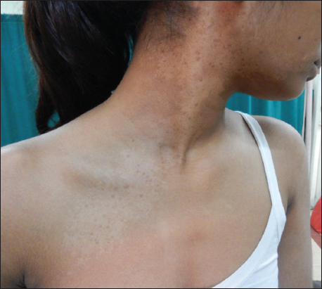
A 17 year girl presented with hypopigmented patch with indented borders on the right side of face and neck which was first noticed by the mother of the child when she was 3 years old. Later on at the age of 5, numerous hyperpigmented punctiform spots appeared exclusively on the hyperpigmented area. With sun exposure the hypopigmented areas neither turned red nor did the skin burn at the site. On local examination, a single ill defined hypopigmented macule of size of about 25 X 8 centimeters was present at the angle of mouth on right side, further extending to lateral side of right ear, right angle of jaw, lateral right side of neck to about 6 centimeters below the clavicle. It was irregular in shape with serrated irregular margins. The surface was smooth and had multiple oval dark brown coloured macules of 1 to 4 mm in size (Fig. 1). On diascopy the margin of the hypopigmented lesion remained delineated. Adjacent oral mucosa was normal.
The dermoscopic examination (Fig. 2) showed 1-4 millimeter sized hyperpigmented lesions, with a barely visible pseudonet, leading to the final diagnosis of colocalized nevus depigmentosus and lentigines

DISCUSSION
The commonly used clinical diagnostic criteria for nevus depigmentosus (ND)are as follows [2–4]:
- Leukoderma present at birth or of an early onset
- No alteration in distribution of leukoderma throughout life
- No alteration in texture or change of sensation in the affected area
- Absence of hyperpigmented border
It may be localized, segmental, or systematized [5]. Wood’s lamp examination shows an off-white accentuation in ND (compared to chalky-white in vitiligo). Histopathologically, the numbers of melanocytes are normal or decreased [2,6] but DOPA reactivity is consistently reduced [2]. Melanosomes are usually normal in size, shape, and internal structure [1], but can be decreased in number, heteromorphic, aggregated in melanocytes, or located in membrane bound aggregates [5].
Until now 8 cases of colocalized nevus depigmentosus and lentigines have been reported [2–4,6,7,8]. The theory of twin spots does not apply to colocalized nevus. Two hypotheses were suggested for the colocalized nevus [8]. The first one, which applies to larger lesions such as syndrome of Ito, hypothesizes that a mutational event occurring in the first 4 weeks of life when the embryo is a single developmental field, leads to a polytopic malformation, namely more associated malformations. In our case, which is characterized by small nevi, the mutation affecting the same melanocytic function, is the more probable hypothesis [8] which is responsible for nevus depigmentosus. This mutation is followed by a reverse mutation of the gene involved in the pigmentation. The latter could restore the pigmentation incompletely. Thus, colocalization of lentigines can be regarded as a different form of repigmentation resulting from reverse mutation in one of the genes involved in pigmentation [2].
Consent
The examination of the patient was conducted according to the Declaration of Helsinki principles.
REFERENCES
1. Lesser E, Ziemssen HV, Hanbuchder Hautkrankheiten 1884; 2nd ed. Leipzig: Vogel; 183-
2. Shim JH, Seo SJ, Song KY, Hong CK, Development of multiple pigmented nevi within segmental nevus depigmentosusJ Korean Med Sci 2002; 17: 133-6.
3. Bolognia JL, Lazova R, Watsky K, The development of lentigines within segmental achromic neviJ Am Acad Dermatol 1998; 39: 330-3.
4. Khumalo NP, Huson S, Burge S, Development of lentigines within naevoid hypopigmentationBr J Dermatol 2001; 144: 188-9.
5. Bhagwat PV, Kudligi C, Odugoudar SG, Krishna S, Pigmented nevi within segmental nevus depigmentosus – a rare caseJ Pak Associat Dermatol 2015; 25: 76-7.
6. Alkemade H, Juhlin L, Unilateral lentiginosis with nevus depigmentosus on the other sideJ Am Acad Dermatol 2000; 43: 361-3.
7. Jagia R, Mendiratt V, Koranne RV, Sardana K, Bhushan P, Solanki RS, Colocalized nevus depigmentosus and lentigines with underlying breast hypoplasia: A case of reverse mutation?Dermatol Online J 2004; 10: 12-
8. Baba M, Akcali C, Seckin D, Happle R, Segmental lentiginosis with ipsilateral nevus depigmentosus: another example of twin spotting?Eur J Dermatol 2002; 12: 319-21.
Notes
Source of Support: Nil,
Conflict of Interest: None declared.
Comments are closed.