A case of pityriasis rosea of vidal accompanied by neurofibromatosis type 1
Ahu Yorulmaz1, Ferda Artuz1, Sezer Kulacoglu2, Elif Sen2
1Ankara Numune Research and Education Hospital, Department of Dermatology, Ankara, Turke, 2Ankara Numune Research and Education Hospital, Department of Pathology, Ankara, Turkey
Sir,
A 23-year-old woman came to our outpatient clinic with a two-weeks history of pruritic reddish lesions on her armpits and groins. Her past medical history was consistent with neurofibromatosis type 1 (NF-1), for which she was under routine follow-up. Upon dermatological examination we observed multiple erythematous to violaceous coalescent and infiltrative plaques with central clearing which were annular and polycyclic in shape on bilateral proximal femoral regions (Figs 1–3) and erythematous papules tending to merge into smaller plaques on left axillary area (Fig. 4). Dermatological examination also disclosed several café-au-lait macules, one of which was greatest in diameter on left lateral brachial region and axillary freckling which was more marked on the left side (Fig. 4). A lesional skin biopsy from erythematous plaques revealed extravasation of red blood cells in dermis, dermal perivascular lymphocytic infiltration and focal spongiosis in the epidermis (Figs 5–7). Based on history, clinical and histopathological findings a diagnosis of pityriasis rosea circinata et marginata of Vidal (PRV) accompanied by NF-1 was made. The patient was prescribed topical corticosteroids and at the one month follow up visit, the lesions were significantly improved.
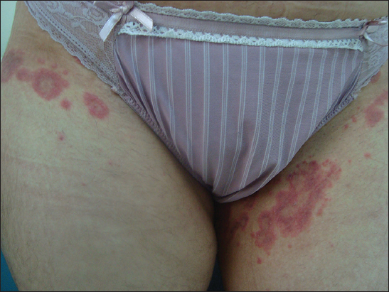
Figure 1: Erythematous to violaceous papules and confluent annular, polycyclic plaques with central clearing
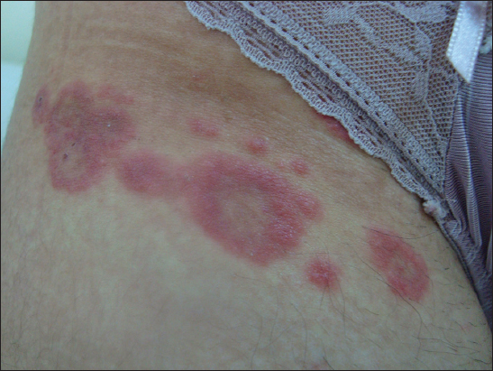
Figure 2: Coalescent plaques with indurated borders and paler central areas
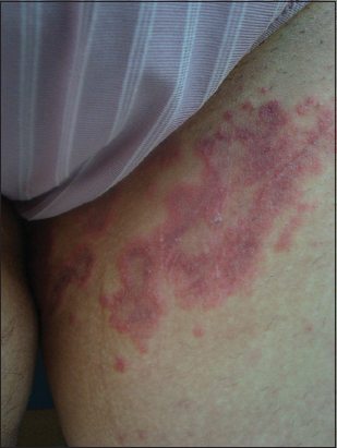
Figure 3: Confluent plaques with central paling and a fine scale in the middle of the largest plaque

Figure 4: Erythematous papules merging into plaques, a large sized café-au-lait macule and axillary freckling
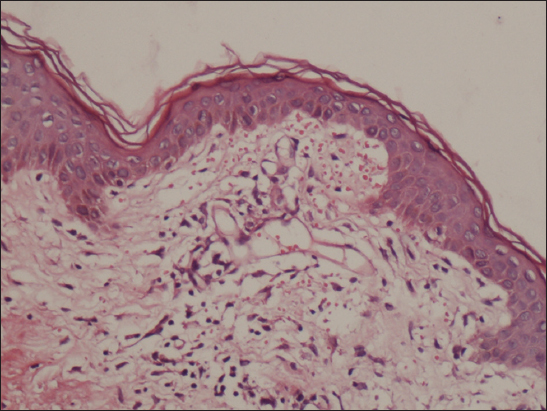
Figure 5: Erythrocyte extravasation within the dermis and superficial dermal perivascular infiltration, (H&E ×200)
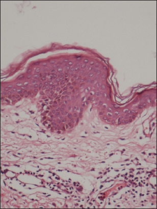
Figure 6: Focal spongiosis in the epidermis (H&E ×200)
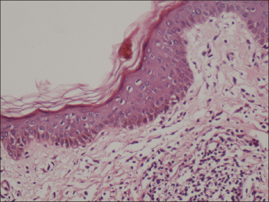
Figure 7: Small foci of parakeratosis and dermal perivascular lymphocytic infiltration, (H&Ex200)
Prior to the study, patient gave written consent to the examination and biopsy after having been informed about the procedure.
Pityriasis rosea (PR) is a self-limited, acute exanthem of uncertain etiology mostly affecting children and young adults [1]. NF-1 is a common genodermatosis characterized by café-au-lait macules, neurofibromas, freckling in the axillae and groin, pigmented iris hamartomas, and skeletal abnormalities. The clinical spectrum of NF is broad and the diagnosis is made on the basis of clinical manifestations [2]. On the other hand, PR has several clinical variants, each with a distinct presentation of morphology, configuration and distribution pattern of the lesions. PRV is an uncommon variant of PR, characterized by large oval or annular plaques around the axilla and / or groins. Affecting exclusively axilla and groin, PRV must be considered in the differential diagnosis of dermatophytosis, secondary syphilis and invers psoriasis [1,3,4]. Herein, we report a case of 23-year-old woman with PRV accompanied by NF Type 1.
CONSENT
The examination of the patient was conducted according to the Declaration of Helsinki principles.
REFERENCES
1. Sterling JC, Burns T, Breathnach S, Cox N, Grittiths C, Virus InfectionsRook’s Textbook of Dermatology 2010; 8th ed. Oxford: Wiley-Blackwell; 33.78-34.1.
2. Tonsgard JH, Clinical manifestations and management of neurofibromatosis type 1Semin Pediatr Neurol 2006; 13: 2-7.
3. Zawar V, Godse K, Annular groin eruptions: pityriasis rosea of vidalInt J Dermatol 2011; 50: 195-7.
4. Zawar V, Kumar R, Multiple recurrences of pityriasis rosea of Vidal: a novel presentationClin Exp Dermatol 2009; 34: e114-6.
Notes
Source of Support: Nil,
Conflict of Interest: None declared.
Comments are closed.