Soft tissue chondroma: a rare tumor presenting as a cutaneous nodule
Dibakar Podder, Vidya Monappa, Prashanth Shetty
Department of Pathology, Kasturba Medical College, Manipal University, Manipal – 576104, Karnataka, India
ABSTRACT
Soft tissue chondroma (STC), also known as extraskeletal chondroma or chondroma of soft parts is a benign cartilaginous tumor which arise de novo from soft tissue. Also, it is an extremely rare entity predominantly involving extremities, especially fingers. A 26 year old male presented with 3 year history of swelling in left index finger. On local examination a hard 2 × 2 cm swelling was seen over the volar aspect of left 2nd proximal phalanx. Swelling was mobile on contraction of tendons. X-ray showed a soft tissue shadow on volar aspect of left second proximal phalanx. Histopathology showed a well encapsulated, hypo cellular nodule composed of benign chondrocytes surrounded by hyaline chondroid matrix. Nuclear pleomorphism, mitosis or necrosis was not seen. Based on radiological and histopathological findings a diagnosis of STC was made. STC should be considered in patients with slow growing, soft tissue masses.
Key words: Chondroma; Soft tissue; Extraskeletal
INTRODUCTION
STC, also known as extraskeletal chondroma or chondroma of soft parts and is a benign cartilaginous tumor. It commonly affects soft tissues of hands and feet [1]. Fingers are most commonly involved followed by hands, toes, feet and trunk. It can also be seen in dura, larynx, pharynx, oral cavity, skin, parotid gland and fallopian tube [2].
CASE REPORT
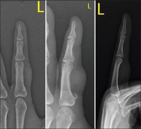
A 26 year old male presented with 3 year history of cutaneous swelling left index finger. No significant past history or history of trauma was present. On local examination a hard 2 × 2 cm swelling seen over the volar aspect of left 2nd proximal phalanx. Swelling was mobile on contraction of tendons. X-ray showed a soft tissue shadow on volar aspect of left second proximal phalanx. The underlying bone was free (Fig. 1). Grossly, the excised specimen consisted of a single nodular tissue bit with dimensions of 1.5 × 1.5 × 1 cm. Cut section was glistening white. Microscopically, a well encapsulated hypocellular nodule was seen composed of benign chondrocytes surrounded by hyaline chondroid matrix. Nuclear pleomorphism, mitosis or necrosis was not seen (Fig. 2). A diagnosis of soft tissue chondroma was made based on clinical, radiological and histopathological findings. The patient has been on regular follow-up (4 months) with no evidence of local recurrence.

The patient’s informed consent was obtained.
Prior to the study, patient gave written consent to the examination and biopsy after having been informed about the procedure.
DISCUSSION
Chondroma is a benign cartilaginous tumor. When it arises in the medullary cavity, it known as enchondroma, which is an extremely common bone tumor. When it arises from soft tissue without attachment to underlying bone, it is known as Soft tissue chondrom [3,4].
STC was first described by Baumuller in 1883 and since then around 200 cases had been reported in the world literature [5]. They commonly arise as painless slow growing swelling in the extremities especially hands and feet. It frequently affects adults of 30-60 years of age and rarely occurs in children [6]. The nodule can be associated with pain or cause nail deformity, depending upon the localization [5]. Radiologically, the tumor is well demarcated and does not involve bone. Ring-like, curvilinear calcification can be seen. Rarely, calcification may be absent contributing to missing the diagnosis of chondroma [7]. The present case did not show evidence of calcification.
Grossly STCs are well circumscribed round to oval masses rarely exceeding 3 cm in greatest diameter. Sometimes it may be friable with cystic changes. Histologically, well circumscribed tumor with lobules of mature chondrocytes or chondroblasts surrounded by hyaline matrix is seen. Less commonly focal fibrosis (fibrochondroma), ossification (osteochondroma) or myxoid change (myxochondroma) can be seen [8,9]. Malignant transformation has not been reported in the literature. Immunohistochemically, the tumour cells are positive for vimentin, S100 and negative for epithelial and myoepithelial markers [10].
Clonal chromosomal abnormalities have been detected in some extraskeletal chondromas including monosomy 6, trisomy 5 and rearrangements of chromosome 11 [11,12]. The histological differential diagnosis includes calcifying aponeurotic fibroma, tumoral calcinosis, periosteal or juxtacortical chondroma, synovial chondromatosis, extraskeletal myxoid chondrosarcoma and chondroid syringoma [13]. Calcifying aponeurotic fibroma occurs in young patients and predominantly involves hands. Microscopically short bar like foci of cartilaginous metaplasia surrounded by infiltrating fascicles of fibromatosis-like plump fibroblasts is characteristic. Tumoral calcinosis mimics heavily calcified chondroma but lacks cartilage and shows histocytic reaction in response to the calcified material. Synovial chondromatosis differs from extraskeletal chondroma by its occurrence in large joints. It is characterized by formation of numerous metaplastic cartilaginous or osteocartilaginous nodules of varying sizes attached to the synovial membrane of the joint, tendon sheath or extra-articular bursa. Microscopically cartilaginous masses just beneath the thin lining of the synovial membrane are seen [14]. Extraskeletal myxoid chondrosarcoma can mimic myxoid variant of STC. However, chondroma is smaller, well defined and less cellular with cells being better differentiated. Chondroid syringoma will show eccrine ducts and glands surrounded by myxoid matrix with cartilaginous differentiation.
CONCLUSION
STC is an extremely rare, slow growing, benign cartilaginous tumor which commonly arises from soft tissue of hands and feet. Complete surgical excision is adequate treatment. Recurrence is uncommon and malignant transformation is rare. STC should be considered in patients with slow growing, soft tissue masses.
CONSENT
The examination of the patient was conducted according to the Declaration of Helsinki principles. Written informed consent was obtained from the patient for publication of this article and any accompanying images.
REFERENCES
1. Chung EB, Enzinger FM, Chondroma of soft partsCancer 1978; 41: 1414-24.
2. Weiss SW, Goldblum JR, Cartilaginous soft tissue tumorsEnzinger and Weiss’s. Soft tissue tumors 2008; 5th Edition. China: 1017-23.
3. Anthouli-Anagnostopoulou FA, Papachristou G, Extraskeletal chondroma, a rare soft tissue tumor. Case reportActa Orthop Belg 2000; 66: 402-4.
4. DelSignore JL, Torre BA, Miller RJ, Extraskeletal chondroma of the hand. Case report and review of the literatureClin Orthop Relat Res 1990; 254: 147-52.
5. Ishii T, Ikeda M, Oka Y, Subungal extraskeletal chondroma with finger nail deformity: case reportJ Hand Surg Am 2010; 35: 296-9.
6. Hondar Wu HT, Chen W, Lee O, Chang CY, Imaging and pathological correlation of soft-tissue chondroma: a serial five-case study and literature reviewClin Imaging 2006; 30: 32-6.
7. Zlatkin MB, Lander PH, Begin LR, Hadjipavlou A, Soft-tissue chondromasAJR Am J Roentgenol 1985; 144: 1263-7.
8. Gulati Y, Maheshwari A, Sharma V, Mattoo R, Arora D, Gupta N, Extraskeletal osteochondroma of the thigh: a case reportActa Orthop Belg 2005; 71: 115-8.
9. Sheff JS, Wang S, Extraskeletal osteochondroma of the footJ Foot Ankle Surg 2005; 44: 57-9.
10. Aslam MB, Haqqani MT, Extraskeletal chondroma of parotid glandHistopathology 2006; 48: 465-7.
11. Dal Cin P, Qi H, Sciot R, Van den Berghe H, Involvement of chromosomes 6 and 11 in a soft tissue chondromaCancer Genet Cytogenet 1997; 93: 177-8.
12. Bridge JA, Bhatia PS, Anderson JR, Neff JR, Biologic and clinical significance of cytogenetic and molecular cytogenetic abnormalities in benign and malignant cartilaginous lesionsCancer Genet Cytogenet 1993; 69: 79-90.
13. Gungor S, Kamali G, Canat D, Gokdemir G, Soft tissue chondroma of the index finger: clinical, histological and radiological findings in a unique caseDermatol Online J 2013; 19: 18176.
14. Polster JM, Evans P, Schils J, Sundaram M, Radiologic case study. Synovial chondromatosis of the pisotriquetral jointOrthopedics 2005; 28: 1130.
Notes
Source of Support: Nil
Conflict of Interest: None declared.
Comments are closed.