Our Dermatol Online. 2012; 3(3): 202-205
Date of submission: 26.03.2012 / acceptance: 23.04.2012
Conflicts of interest: None
IMMUNOLOGIC FINDINGS IN CENTRAL CENTRIFUGAL CICATRICIAL ALOPECIA
Ana Maria Abreu Velez1, A. Deo Klein2, Michael S. Howard1
1Georgia Dermatopathology Associates, Atlanta, Georgia, USA
2Statesboro Dermatology, Statesboro, Georgia, USA
Corresponding author: Ana Maria Abreu Velez, MD PhD e-mail:abreuvelez@yahoo.com
How to cite an article: Abreu Velez AM, Klein AD, Howard MS. Immunologic findings in central centrifugal cicatricial alopecia. Our Dermatol Online 2012; 3(3): 202-205.
Abstract
Introduction: Premature desquamation of the inner root sheath is described as a defining histologic feature of follicular degeneration syndrome/central centrifugal cicatricial alopecia; moreover, the immunological features of this disease are not well established.
Case report: A 46-year-old African American female was evaluated for an asymptomatic scarring alopecia after using several chemicals on her hair. The clinical examination revealed visible, well defined patches of hair loss.
Methods: Biopsies for hematoxylin and eosin examination, as well as for direct immunofluorescence and immunohistochemistry analysis were performed. We evaluated molecules involved in signaling of growth factor pathways (e.g. the Akts), specifically VEGF and Oct-4 to investigate involvement of these molecules in this disease. Hematoxylin and eosin staining demonstrated histopathologic findings of premature desquamation of the inner root sheath and eccentric thinning of the follicular epithelium, supporting the diagnosis of central centrifugal cicatricial alopecia. Direct immunofluorescence revealed strong depositions of IgG, Complement/C3 and fibrinogen around the multiple hair follicles and their supply vessels. Immunohistochemistry staining of the base of the hair follicle was seen with fibrinogen and Oct-4 antibodies. Immunohistochemistry also demonstrated increased expressions of VEGF around supply vessels of the hair follicle, as well as some overexpression of anti-human Akt-pS473 phosphorylation site specific antibody.
Conclusions: Our immunologic findings suggest that the etiology of centrifugal cicatricial alopecia includes not only hair traction, but also a possible reactive immune response.
Key words: central centrifugal cicatricial alopecia; embryonic stem factor; Oct-4; follicular degeneration syndrome; immunofluorescence; Akt
Abbreviations and acronyms:
Immunohistochemistry (IHC), hematoxylin and eosin (H&E), central centrifugal cicatricial alopecia (CCCA), vascular endothelial growth factor (VEGF), octamer-binding transcription factor 4 (Oct-4).
Introduction
Follicular degeneration syndrome, also known as central centrifugal cicatricial alopecia is a form of scarring alopecia which is most often clinically first visible as a well defined patch of diffuse hair loss [1-3]. The affected region frequently, although not always, extends centrifugally from the scalp vertex. The disease area may gradually expand in size with time [1-3]. Skin biopsies show that central centrifugal cicatricial alopecia involves inflammation of the affected hair follicles and early desquamation of the hair follicle internal root sheath [1-3].
Case report
A 46-year-old African American female was evaluated for alopecia following several chemical treatments on her hair. The clinical examination revealed visible, well defined patches of diffuse hair loss. The affected region extended centrifugally from the scalp vertex. The patient denied any other systemic disease, and multiple laboratory tests were negative including antinuclear antibodies (ANAs). Biopsies for hematoxylin and eosin (H&E) examination, as well as for direct immunofluorescence (DIF) and immunohistochemistry (IHC) analysis were performed.
Direct immunofluorescence (DIF):
In brief, skin cryosections were prepared, and incubated with multiple fluorochromes as previously reported [3-8]. We utilized normal skin as a negative control, obtained from aesthetic plastic surgery patients. We utilized FITC conjugated Immunoglobulins A, E, G, M and Complement/C3 from Dako (Carpinteria, California, USA). We also tested for molecules involved in signaling by the growth factors pathway (e.g. the Akts), specifically VEGF and embryonic stem factor marker Oct-4 (Cell Signaling Technology, Danvers, Massachusetts, USA) to determine involvement of these molecules in this disease. We utilized Alexa 647 (Invitrogen, Carlsbad, California, USA) as a secondary DIF antibody fluorochrome.
Immunohistochemistry (IHC):
IHC was performed as previously described. We utilized antibodies against human monoclonal rabbit anti-human Akt-pS473 phosphorylation site specific antibody and monoclonal mouse anti-human VEGF; these antibodies were also obtained from Dako [3-8].
Results
Microscopic description:
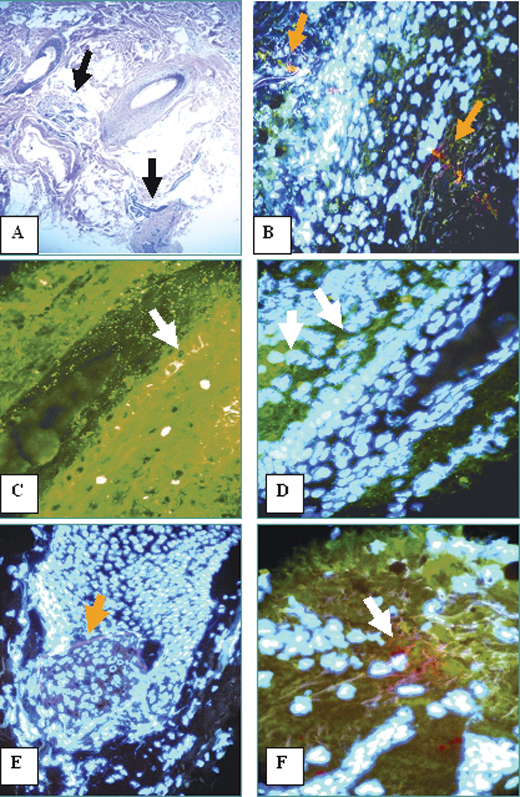
Hematoxylin and eosin (H&E) staining demonstrated histopathologic findings of premature desquamation of the inner root sheath and eccentric thinning of the follicular epithelium, supporting the diagnosis of central centrifugal cicatricial alopecia (CCCA). The overall anagen:telogen ratio appeared within normal limits. Focal perifollicular, concentric fibrotic scarring was observed, approximating five (5) per cent of the biopsy area. A mild to moderate perivascular and peri-infundibular inflammatory infiltrate was also noted, with attendant loss of hair follicular units (Fig. 1, 2). The infundibular epithelium was also noticeably atrophic. Focal dermal follicular stelae scars were identified. A Verhoeff elastin special stain confirmed the extent of scarring within the dermis (Fig. 2). Direct immunofluorescence revealed strong depositions of Complement/C3, fibrinogen and some IgG around the hair follicles and surrounding supply vessels. The immune reactivity colocalized with the presence of Oct-4. Immunohistochemistry demonstrated increased some overexpression of VEGF and anti-human Akt-pS473 phosphorylation site specific antibody in supply vessels around the hair follicles.
Discussion
Follicular degeneration syndrome was also previously known as central progressive alopecia, or hot comb alopecia. The entity was first identified in African-American women, and thought to be secondary to heat of hot combs and oil pomades [1-3,9]. It was originally thought that the oils applied to the hair were heated by the hot comb. The liquid oil was then believed to stream down the hair fiber into the hair follicle opening and irritate the skin, causing inflammation around the upper hair follicle [1-3,9]. Nevertheless, it is now known that although hot combing might elicit follicular degeneration syndrome in some individuals, the disorder may also occur in the absence of any cosmetic procedure. The clinical differential diagnosis of follicular degeneration syndrome is extensive and includes the following disorders: lichen planopilaris, frontal fibrosing alopecia, fibrosing alopecia in a pattern distribution, central pseudopelade of Brocq, traction alopecia, secondary systemic scarring alopecia (e.g lupus), trichotillomania, chemotherapy alopecia, alopecia mucinosa, keratosis pilaris atrophicans, mycosis fungoides, perifolliculitis capitis abscedens et suffodiens, tufted hair folliculitis, acne keloidalis, acne necrotica, erosive pustular dermatosis of the scalp, pressure alopecia, lipedematous alopecia, and senescent alopecia [1-3,9]. Our immunofluorescence and immunohistochemistry studies reveal that some immune response against the hair follicles and/or their vessels is present. The autoantibodies colocalize with Oct-4, previously characterized as a protein in humans encoded by the POU5F1 gene, and critically involved in the self-renewal of undifferentiated embryonic stem cells [10]. We thus speculate that the hair follicle embryonic cell population could be altered in this disease, although we have analyzed only one case. Activated Akt phosphorylated protein (also known as Akt2 and protein kinase B; antibody Akt-pS473) functions as an important regulator of various cell processes including apoptosis, proliferation, differentiation and metabolism. We found that this protein seems to be overexpressed around the hair shafts and vessels [11]. Akt2 is a critical downstream effector of PI3-kinase, which mediates signal transduction when initiated by a variety of stimuli including hormones, growth factors and cytokines [11]. Some authors have shown that Akt2 is a determinant of postnatal hair follicle development [11]. Thus, we suggest that more cases of this disease should be compiled to study the significance and colocalization of the immune response around the hair follicles, utilizing our study markers and other antibodies.
|
Figure 1. a. The H&E demonstrates the presence of fibrosing alopecia and atrophy of multiple hair follicles (black arrows) (100X). In b. DIF double staining with FITC conjugated anti-human fibrinogen (green staining) and Alexa 647 conjugated Oct-4 (red staining) around several vessels surrounding a hair follicle. The follicle nuclei are counterstained with Dapi (light blue/white). In c. DIF using FITC conjugated complement/C3 (green staining; white arrow) that shows positivity around a hair shaft and also in some cells around the hair follicle. In d. similar to 1c. but using Dapi nuclear counterstaining. In e. note positivity in the hair bulb with Oct-4 (red staining; yellow arrow). In f. note positivity around hair follicle supply vessels using Oct-4 (red staining; white arrow).
|
|
Figure 2. a. Shows a Verhoeff elastin stain, with the red arrow pointing to an atrophic hair follicle. In Figure b. H&E staining shows atrophic hair follicles. In c. and d. VEGF and Akt-pS473 are overexpressed by IHC at the hair follicle and surrounding vessels, respectively (dark staining; red arrows).
|
REFERENCES
1. Horenstein MG, Simon J: Investigation of the hair follicle inner root sheath in scarring and non-scarring alopecia. J Cutan Pathol. 2007; 34: 762-768. 2. Elston DM, Ferringer T, Dalton S, Fillman E, Tyler W: A comparison of vertical versus transverse sections in the evaluation of alopecia biopsy specimens. J Am Acad Dermatol. 2005; 53: 267-272. 3. Abreu-Velez AM, Klein AD, Howard MS: Survivin, p53, MAC, Complement/C3, fibrinogen and HLA-ABC within hair follicles in central and centrifugal cicatricial alopecia. N Am J Med Sci. 2011; 3: 292-295. 4. Abreu-Velez AM, Girard JG, Howard MS: Antigen presenting cells in the skin of a patient with hair loss and systemic lupus erythematosus. 2009; 1: 205-210. 5. Abreu-Velez AM, Smith JG Jr, Howard MS: Activation of the signaling cascade in response to T lymphocyte receptor stimulation and prostanoids in a case of cutaneous lupus. N Am J Med Sci. 2011; 3: 251-254. 6. Abreu-Velez AM, Brown VM, Howard MS: Antibodies to piloerector muscle in a patient with lupus-lichen planus overlap syndrome. N Am J Med Sci. 2010; 2: 276-280. 7. Abreu Velez AM, Dejoseph LM, Howard MS: HAM56 and CD68 antigen presenting cells surrounding a sarcoidal granulomatous tattoo. N Am J Med Sci. 2011; 3: 475-477. 8. Abreu Velez AM, Brown VM, Howard MS: An inflamed trichilemmal (pilar) cyst: Not so simple? N Am J Med Sci. 2011; 3: 431-434. 9. Summers P, Kyei A, Bergfeld W: Central centrifugal cicatricial alopecia – an approach to diagnosis and management. Int J Dermatol. 2011; 50: 1457-1464. 10. Tsai SY, Bouwman BA, Ang YS, Kim SJ, Lee DF, Lemischka IR, et al: Single transcription factor reprogramming of hair follicle dermal papilla cells to induced pluripotent stem cells. Stem Cells. 2011; 29: 964-971. 11. Mauro TM, McCormick JA, Wang J, Boini KM, Ray L, Monks B, et al: Akt2 and SGK3 are both determinants of postnatal hair follicle development. FASEB J. 2009; 23: 3193- 31202.


Comments are closed.