Our Dermatol Online. 2011; 2(3): 141-143
Date of submission: 30.03.2011 / acceptance: 22.04.2011
Conflicts of interest: None
ERYTHEMATODES CHRONICUS PROFUNDUS AS DERMATOLOGY, SURGERY AND COSMETOLOGY PROBLEM
Alendar Faruk, Soskic Samra, Helppikangas Hana, Gavrankapetanovic Alma, Alendar Temeida
Dermatovenerology Department Clinical Center University of Sarajevo, Bosnia & Herzegowina
Corresponding author: Dr. Temeida Alendar e-mail: derm_alendar@hotmail.com
How to cite an article: Alendar F, Soskic S, Helppikangas H, Gavrankapetanovic A, Alendar T. Erythematodes chronicus profundus as dermatology, surgery and cosmetology problem. Our Dermatol Online 2011; 2(3): 141-143.
Abstract
Lupus profundus is a rare skin condition originally described by Kaposi in 1883. It is a clinical variant of LE in which the deep dermis and subcutaneous fat are predominantly affected. A 38-year-old female presented with history of development of well-circumscribed patches of hair loss since 1994 year. In 2004 on the skin of left cheek she noticed a single erythematous plaque on which place later became dint of subcutaneous tissue. Today she has massive defects across whole scalp, right cheek and the upper part of left arm with multiple ulcers(fig.3and4). case reposrt, laboratory examinations, skin biopsy and influence of corticosteroids on further progression /regression of this disease.
Key words: lupus profundus; chloroquin; corticosteroids
Introduction
Lupus profundus is a rare skin condition originally described by Kaposi in 1883 and later by Irgang in 1940. It is a clinical variant of LE in which the deep dermis and subcutaneous fat are predominantly affected. It may occur on its own or in association with discoid lupus erythematosus (DLE) or SLE. In cases associated with SLE, it may precede SLE by several years. In the majority of cases, it tends to have a mild chronic course marked by recurrent nodules or plaques.;
Case Report
A 38-year-old female presented with history of development of well-circumscribed patches of hair loss since 1994. year. The hair loss was rapid and asymptomatic, with no hair re-growth. There was no family history of any autoimmune rheumatic diseases. In 2004. she was first time hospitalised on our clinic. Clinical symptoms were: on the skin of left cheek a single, erythematous plaque (Fig. 1). On left arm two small ulcers size 1 x 2 cm, oval in shape and firm in consistency (Fig. 2). Since than she was treated with chloroquin in dose 250mg per day with gradually reduction, but with no results. Today she have massive defects across whole scalp, right cheek and the upper part of left arm (Fig. 3,4). The lesion, on the left arm in 2004. initially, was about 1 Χ 2 cm 2 in size, oval in shape and firm in consistency. It gradually increased in size to about 8 Χ 5 cm 2, in 20l0. with multiple ulcers colonized with Staphilococcus aureus,and with extensive atrophy of cutaneus and subcutaneous tissue. There was no history of preceding trauma. No history of taking injection. She denied arthralgia, arthropathy, myalgia, fatigue, fever, Raynaud’s phenomenon and gastrointestinal symptoms. She notifies pain in right lumbal area, and the ultrasound showed signs of bilateral nephropathy. The following laboratory tests were done: complete blood cell count, differential cell count, ESR was 25, total serum proteins 86.0, with A:G ratio 1.0,y globulins were increased 0.27. Blood sugar, blood urea, serum creatinine, serum electrolytes, liver function tests and urinalysis were in normal rates. X-ray chest was normal. Rheumatoid factor was normal,but antistreptolysin was 221,0IU/ml. C3 1.090 g/l, C4 0.l65 g/l, ANA positive, Anti ds DNA negative and CiC was increased and his amount was 58.o8IU/ml. Skin biopsy showed lymphocytic panniculitis, hyaline degeneration of the fat, hyaline papillary bodies, and lymphoid nodular structures in the lower dermis and subcutaneous tissue. Result is in high correlation with diagnosis of erytematodes cronicus profundus. In 2010. we treated her with systemic corticosteroid therapy in dose of 60mg per day with gradually reduction of dose. After 2 month of therapy further progression has stoped (Fig. 5,6). Her maintence dose today is 20 mg prednisolon/day.
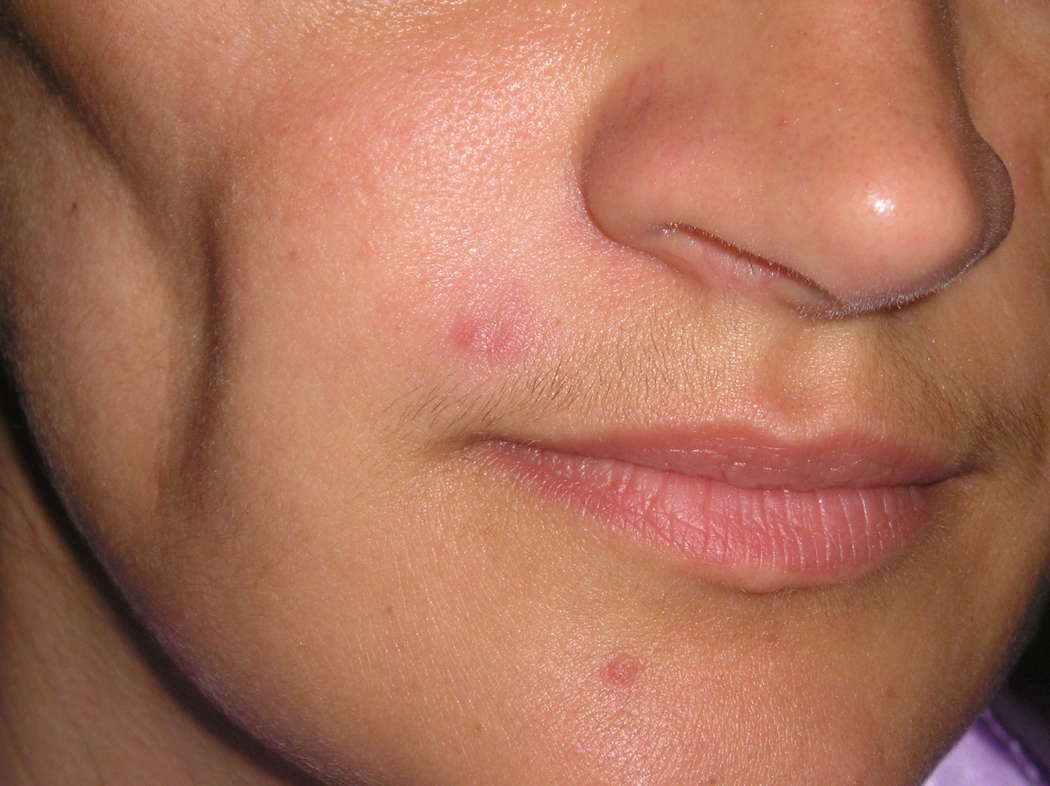 Figure 1. On the skin of left cheek a single, erythematous plaque
|
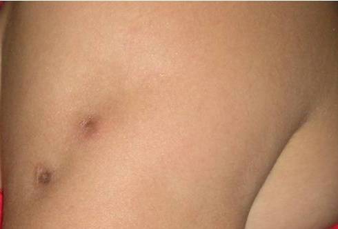 Figure 2. On left arm two small ulcers size 1 x 2 cm, oval in shape and firm in consistency
|
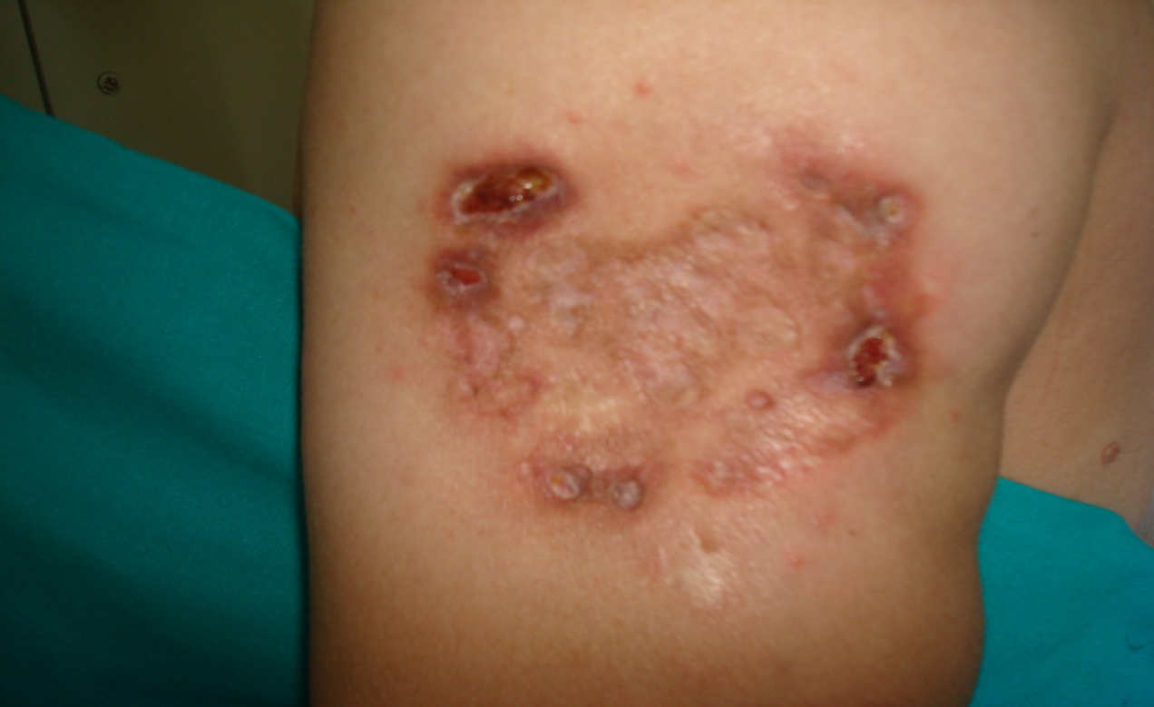 Figure 3. Massive defects across whole the upper part of left arm
|
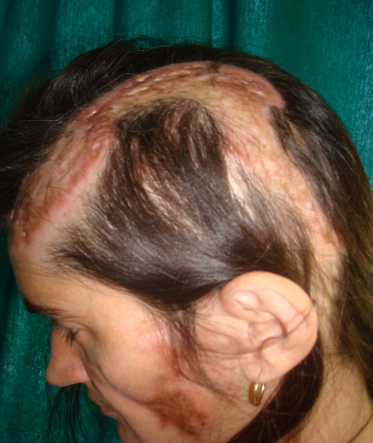 Figure 3. Massive defects across whole scalp, right cheek
|
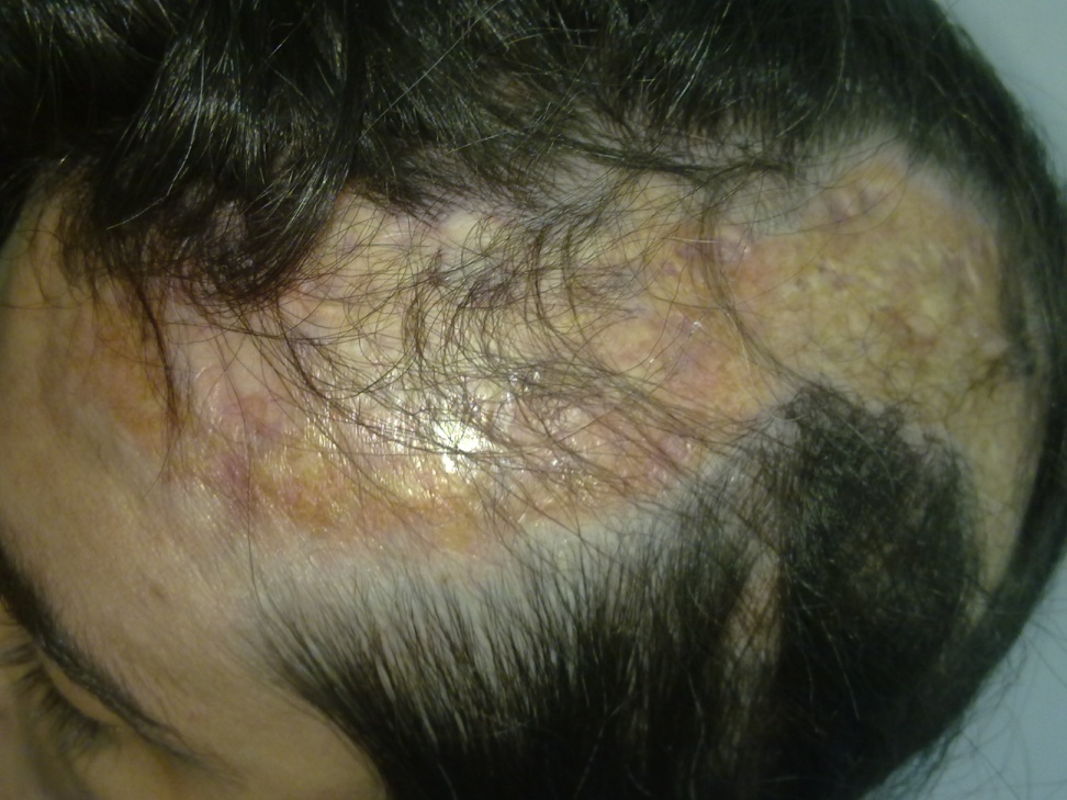 Figure 5. Lesions after 2 month of therapy
|
 Figure 6. Lesions after 2 month of therapy
|
Discussion
Lupus erythematosus profundus or panniculitis is an unusual but distinct clinical variety of lupus erythematosus. The inflammatory reaction in takes place primarily in the deep corium and the subcutaneous tissues leading to deep indurated nodules or sharply defined plaques. The overlying skin usually appears normal but there may be erythema, atrophy, ulceration or poikilodermatous or hyperkeratotic changes. The lesions are most frequent on cheeks but other sites of predilection are face, upper arms, hands, chest, buttocks and thighs. It may develop in association with discoid lupus erythematosus or systemic lupus erythematosus or may occur as an isolated phenomenon. The differential diagnoses include panniculitis due to other connective tissue disorders like dermatomyositis or scleredema and Weber Christian panniculitis or Jessner’s lymphocytic infiltration, lyrnphocytoma cults and sarcoidosis. Lupus panniculitis often responds to treatment with antimalarials, such as hydroxychloroquine (200 mg once or twice a day). Some cases respond to a combination of antimalarials (for example, hydroxychloroquine 200 mg and quinacrine 100 mg daily) when monotherapy is ineffective. Systemic glucocorticoids should be reserved for widespread and resistant lesions. Intralesional glucocorticoids are usually ineffective and may exacerbate the atrophic healing process. Success with dapsone, azathioprine, and thalidomide has been described in isolated case reports. Surgical debridement or resection of individual lesions may be attempted when all other modalities have failed and there is appreciable debilitation.
Conclusion
Progress of Erythematodes profundus is unpredictable. As we can see on example of this patient therapy with cloroquine was insufficient to stop progression of disease. With dose of 60 mg methylprednisolon there was no more further progression.Today, after 8 months of last hospitalisation we have not noticed any progression. Defects on skin with which she needs to live daily, are horrifying, what caused her phsychiatric problem and antidepresive therapy. Today she stay as dermatology, surgery and cosmetology problem.
REFERENCES
1. Tuffanelli DL: Lupus erytheratosus panniculitis (profundus). Arch Dermatol 1971; 103: 231-242.
2. Black MM, Cunliffe WJ: Inflammatory disorders of subcutaneous fat. In: Champion RH, Burton JL, Burns DA, et al., editors. Textbook of Dermatology. London: Blackwell Science LTD; 1998.
3. Braun-Falko O,Wolf HH: Dermatology and venerology, New York; 1997.
4. Sanchez NP, Peters MS, Winkelmann RK: The histopathology of lupus erythematosus panniculitis. J Am Acad Dermatol. 1981; 5: 673-680.
5. Ahmed I, Ahmed D. Lupus erythematosus panniculitis: a unique subset within the lupus erythematosus spectrum. Am J Dermatopathol. 2000; 22: 352.
6. Kündig TM, Trüeb RM, Krasovec M: Lupus profundus/panniculitis. Dermatology. 1997; 195: 99-101.
7. Diaz-Jouanen E, DeHoratius RJ, Alarcon-Segovia D, Messner RP: Systemic lupus erythematosus presenting as panniculitis (lupus profundus) Ann Intern Mes. 1975; 82: 376-379.
8. Masood Q, Manzoor R: Lupus erythematosus profundus (panniculitis) J K Practioner. 1995; 2: 135–136.
Comments are closed.