N Dermatol Online. 2010; 1(2): 19-21
Conflicts of interest: None
CHRONIC MUCOCUTANEOUS CANDIDIASIS – A CASE REPORT
1Hassan Iffat, 2Mashkoor Ahmed, 2Hayat Bhat Mohammad, 1Manaan Qazi, 2Shah Parvaiz Ahmad,
1Masood Qazi
1Deptament of Dermatology, STD, Leprosy Govt. Medical College & Associated SMHS Hospital, Srinagar-Kashmir, India
2Department of Medicine, Govt.Medical College, Associated SMHS Hospital, Srinagar-Kashmir, India
Corresponding author: Dr. Iffat Hassan e-mail: hassaniffat@gmail.com
How to cite this article: Hassan I, Mashkoor A, Hayat Bhat M, Manaan Q, Shah PA, Masood Q. Chronic mucocutaneous candidiasis – a case report. Our Dermatol Online 2010; 1(2): 19-21.
Abstract
Chronic mucocutaneous candidiasis(CMC) is a rare group of overlapping syndromes that have in common a clinical pattern of persistent and diffuse cutaneous or mucosal candidal infections. It is usually associated with multiple endocrine dysfunctions and autoimmune disorders therefore patients needs a complete systemic evaluation. Patients of CMC are also susceptible to other fungal and viral infections due to impaired cell mediated immunity. We report a case of CMC wherein the cutaneous and mucosal lesions were not associated with any systemic disorder. The patient responded to topical clotrimazole.
Key words: Chronic mucocutaneous candidiasis; Clotrimazole
Introduction
Chronic mucocutaneous candidiasis (CMC) is an immune disorder of T-cells characterized by chronic infections with candida involving mucosa, skin and nails [1]. It does not represent a specific disease but rather a presentation of a spectrum of immunologic, endocrinologic and autoimmune diseases. Endocrine dysfunctions are the most common underlying disorder associated with CMC [2]. It is a rare disorder and may become apparent at any time in life but it typically presents before three years of age [3]. CMC is not associated with a high degree of mortality because disseminated invasive candidal infections are rare, however, significant morbidity is associated with persistent skin or mucosal infections, endocrinopathies and other associated autoimmune diseases. We report a case of CMC in a nine month old child.
Case Report
A nine month old female child was brought to us with a history of persistent erosions in perianal area, oral ulcers and annular lesions on both knees. Her parents revealed that the baby was three days old when she started with some eruption in perianal area which increased in size and turned into erosions within a period of two weeks and lesions were persistent. One month back, patient started developing oral ulcers and annular lesions on both knees and swelling of the nail folds of some digits of both hands. The patient was born out of a non–consanguineous marriage and there was no family history of similar disease. On examination ,there was erythema, erosions and oozing from perianal area (fig.1). There were two annular plaques around each knee joint with peripheral erythema and scaling and central clearing (fig.2). The middle and index finger nails of left hand and right thumb nails had paronychia and dystrophy of nail plates (fig.3). The examination of oral cavity revealed small ulcerative lesions on lateral margins of tongue and resorption of tip of the tongue giving a bifid appearance (fig.4). The rest of the cutaneous examination was normal. There was no localized or generalized lymhadenopathy. The general physical and systemic examination was normal. Ophthalmological examination was also noncontributory. Complete blood counts ,liver function tests and kidney function tests were normal. Chest radiography and ultrasonography of abdomen were normal. Thyroid profile, parathyroid hormones, serum calcium, phosphate, uric acid and serum cortisol level were all within normal range. Scrapings from skin lesions and nail plates were subjected to potassium hydroxide examination and culture on Sabouraud’s dextrose agar medium. Candida albicans was isolated from all the three sites. On the basis of history, clinical examination and investigations we made a diagnosis of isolated chronic mucocutaneous candidiasis. The patient was treated with topical clotrimazole and the lesions cleared completely in four weeks.
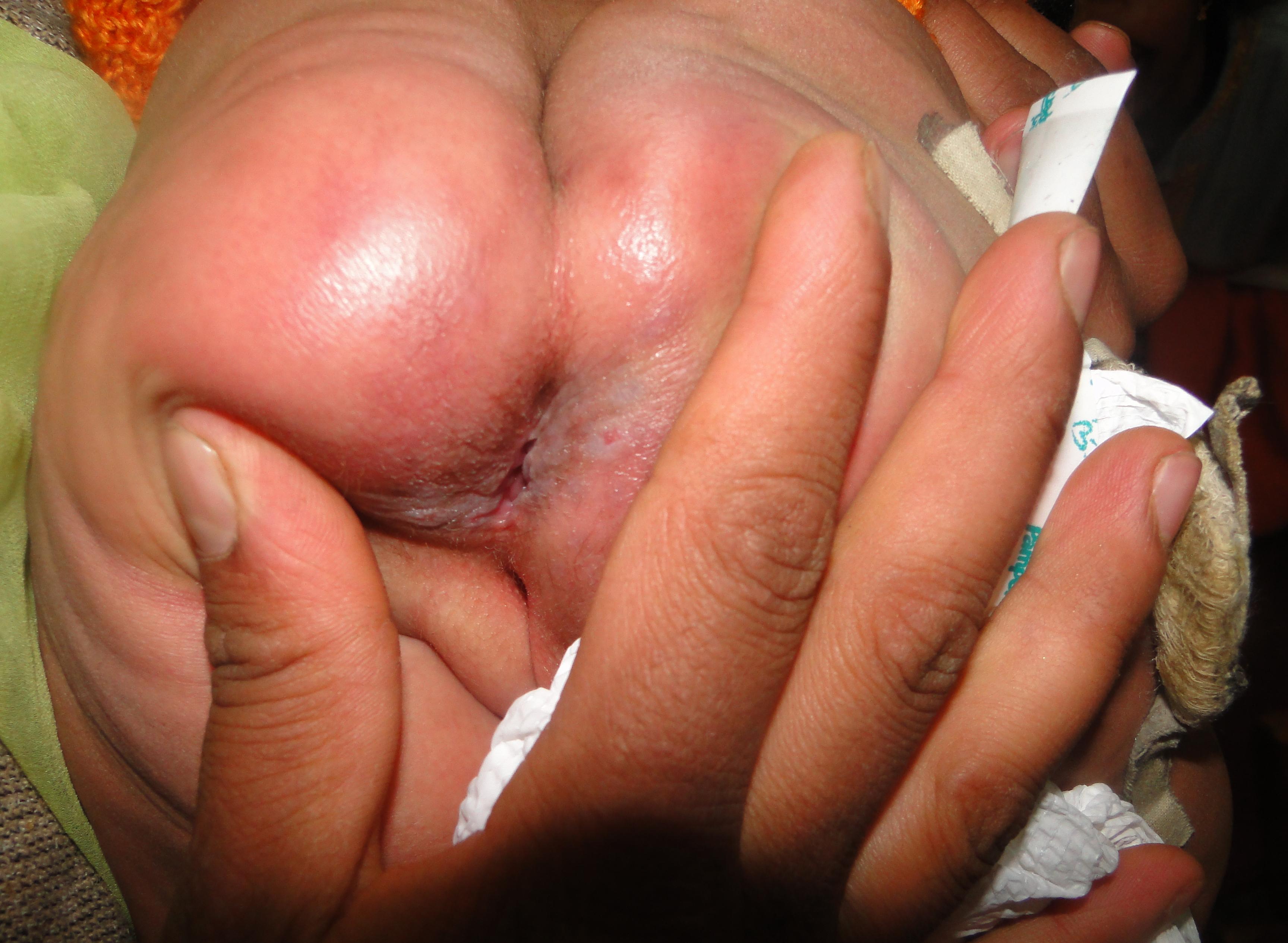 Figure 1.
|
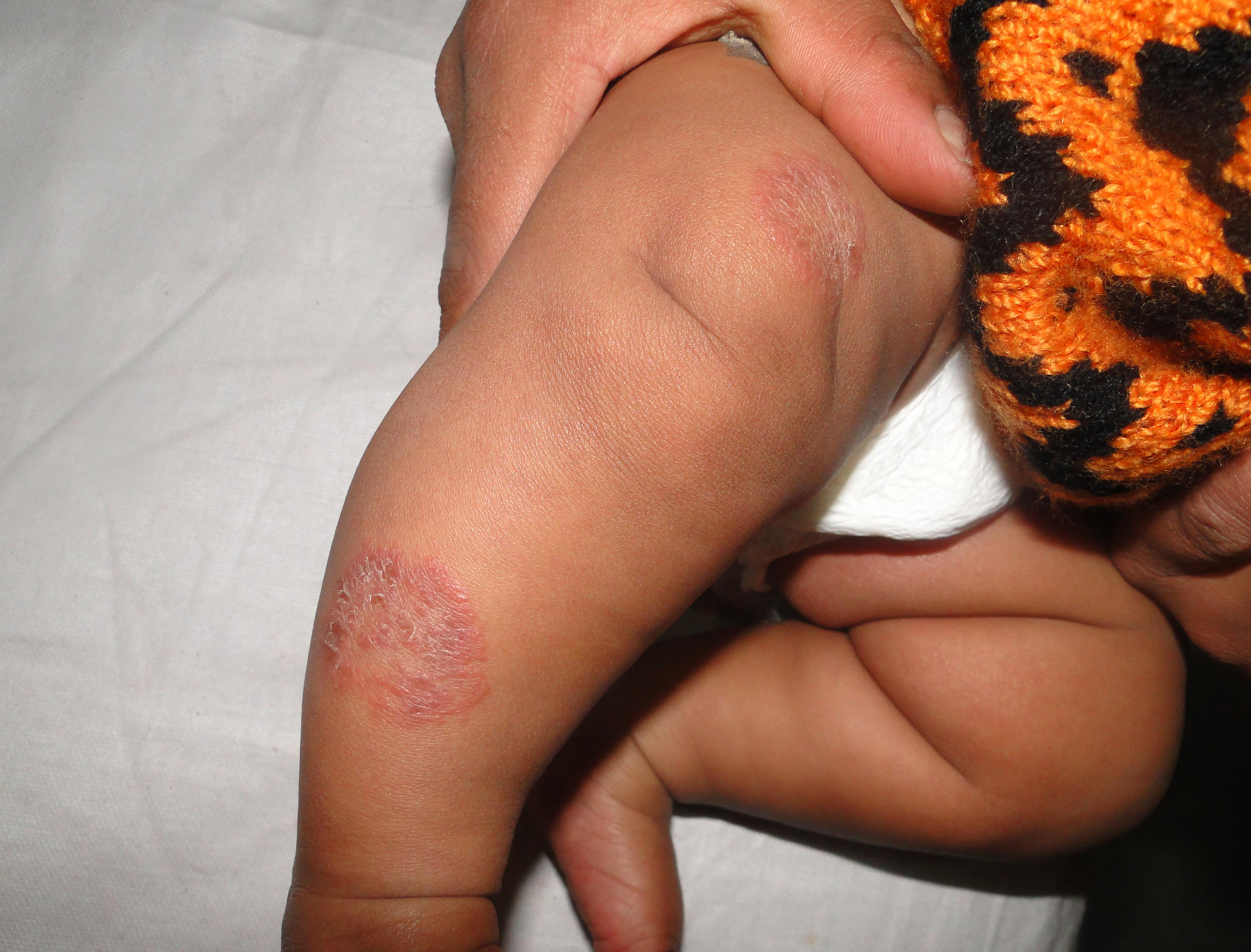 Figure 2.
|
 Figure 3.
|
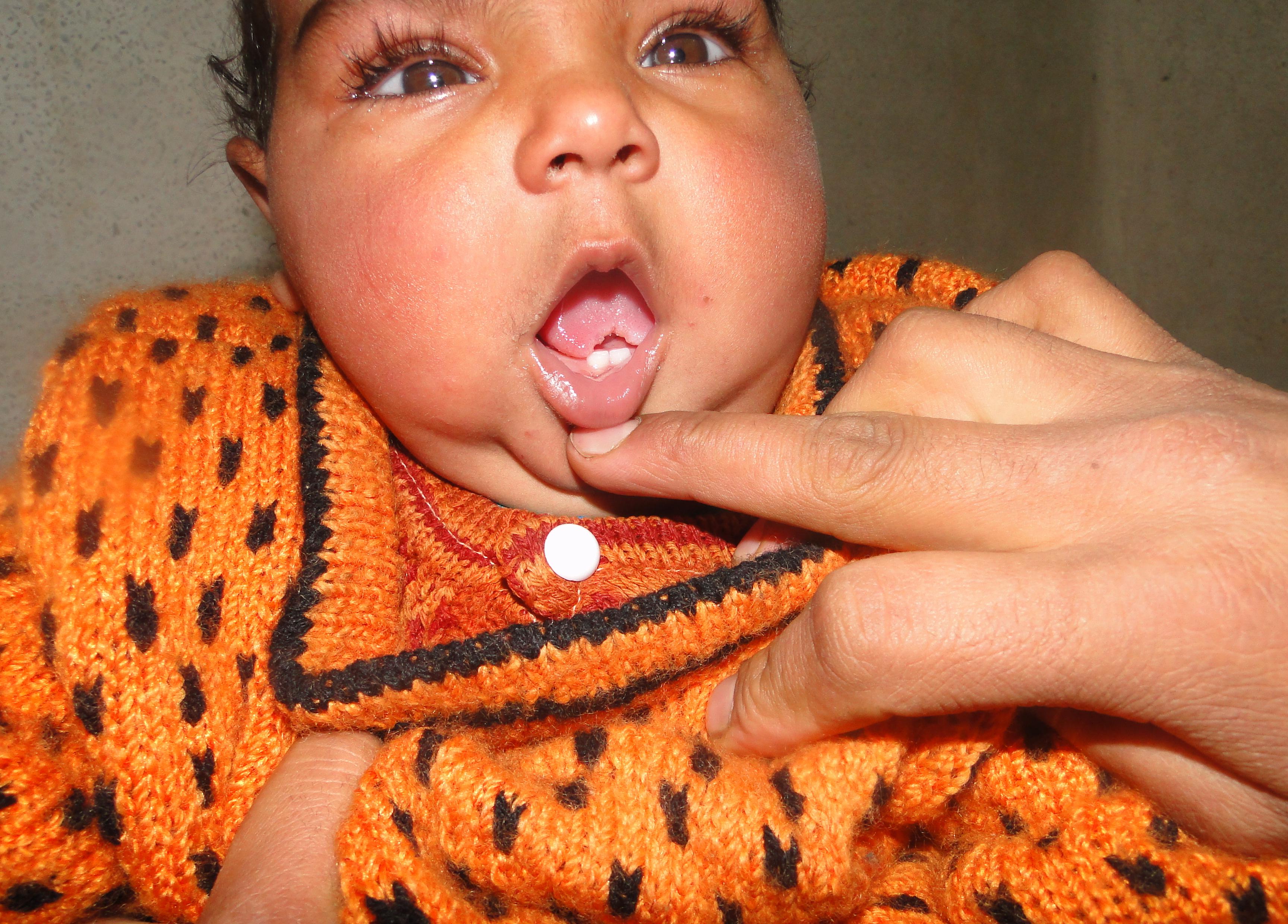 Figure 4.
|
Discussion
Chronic mucocutaneous candidiasis (CMC) is a rare condition characterized by widespread and persistent infection of skin and mucosa caused by candida albicans. CMC is associated with defect in cell mediated immunity. Recent studies suggest that there is alteration in production of cytokines in response to candida antigens. These include decreased interleukin-2 and interferon-gamma levels (Th 1 cytokines) and increased interleukin-10 (Th2 cytokines) [4]. CMC can be limited to skin and mucosa only or it may be associated with polyendocrinopathy, thymoma and KID (keratitis, icthyosis, deafness) syndrome [5]. Hypoparathyroidisim and adrenal failure are the most common endocrine abnormalities associated with CMC [6]. Other infections like dermatophytosis, herpes simplex and disseminated mycobacterium avium infection are also reported to coexist with CMC [7]. Gastrointestinal and hematological dysfunctions are sometimes associated with CMC [7]. In our case only skin ,mucosa and nails were affected and no systemic involvement was detected. There was no predisposing factor for oral candidiasis in our case and the bifid appearance of the tip of the tongue due to persistent infection was an atypical finding in our patient. Current therapy for CMC mainly revolves around prolonged use of antifungal agents especially the azole group [8]. Depending upon the underlying immunologic nature of CMC, various treatments like bone marrow and thymus transplantation or transfusion with white blood cells and candida specific antigens have been attempted [9,10]. In our patient, the lesions cleared completely with topical clotrimazole in four weeks.
Conclusion
We present the case for the unusual clinical presentation in an adult patient (bulky lesions showed a rare anatomical site), having to resort to auxiliary diagnostic methods, and the satisfactory therapeutic response.
REFERENCES
1. Willium J ,Berger D, Timothy G.: Andrews diseases of skin; Clinical Dermatology. Saunders. Elsevier.2006.
2. Coleman R, Hay RJ.: Chronic mucocutaneous candidiasis associated with hypothyroidism; a distinct syndrome.Br J Dermatol 1997; 136: 24-29.
3. Kirkpatrick C H.: Chronic mucocutaneous candidiasis. J Am Acad Dermatol. 1994; 31: 14-17.
4. Lilic D, Cant AJ, Abinum JE, Spickett GP.: Chronic mucocutaneous candidiasis: Altered antigen stimulated IL- 2,IL-4, IL-6 and interferon gamma production. Clin Exp Immunol. 1996; 105: 205-212.
5. Kirkpatrick C H.: Chronic mucocutaneous candidiasis in Candidiasis:pathogenesis, diagnosis and treatment; Raven Press Ltd. New York. 1993.
6. Blizzard R M, Gibbs J H.: Candidiasis: studies pertaining to its association with endocrinopathies and pernicious anaemia. Pediatrics 1968; 31: 231-237.
7. Chipps BE, Saulsburg FT, Hsu SH, Winkelstein JA.: Non-candidal infections in children with Chronic mucocutaneos candidiasis. Johnhopkin Med J. 1979; 144: 175-179.
8. Mobacken H, Moberg S.: Ketoconazole: treatment of 13 patients with chronic mucocutanious candidiasis: A prospective 3 year trial. Dermatologica. 1986; 173: 229- 236.
9. Aiuti F, Businco L, Gatti R.: Reconstitution of T-cell disorders following thymus transplantation. Birth Defects Orig Artic Ser. 1975; 11: 370-376.
10. Buckley RH, Lucos ZJ, Hattler BJ, Amos BD.: Defective cellular immunity associated with chronic mucocutaneous candidiasis: immunological reconstitution by allogenic bone marrow .Clin Exp Immunol. 1968; 3: 153-169.
Comments are closed.