Follicular pityriasis versicolor-Rare variant of a common dermatological disease
Tasleem Arif1,2, Shagufta Rather3
Sir,
Pityriasis versicolor is a superficial fungal infection of the skin caused by yeast of the genus Malassezia. It is characterized by hypopigmented or hyperpigmented macules and patches usually involving the trunk [1]. The variants of pityriasis versicolor include hypochromic, hyperchromic, combination of hypo-hyperchromic, erythematous, circinate, atrophic and acral [2–4]. However, the follicular variant of pityriasis versicolor has been rarely reported. In this article we describe a young female with the follicular variety of this common skin disease.
CASE REPORT
A 30 year old female presented with a one month history of multiple light brownish colored macules over the neck, trunk and proximal limbs. It was associated with mild pruritis. She denied any previous history of similar complaints. On examination, there were multiple light brownish macules present in a follicular distribution over neck, chest, abdomen, back, proximal arms and thighs (Figs 1 and 2). On stretching the affected skin, the scaling became prominent showing the positive Zireli’s sign. Hair, nail and mucous membranes were normal. KOH examination of skin scrapings showed the presence of multiple short hyphae and spores confirming fungal infection. A skin biopsy from the lesion over back showed small 2-4 µm spores in the horny layer of the epidermis. The dermis contained mild inflammatory infiltrate. The adnexal structures including the hair follicles were unremarkable.
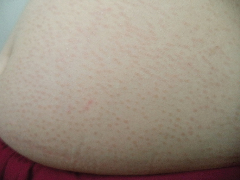
Figure 1: Multiple light brownish macules in a perifollicular distribution over abdomen
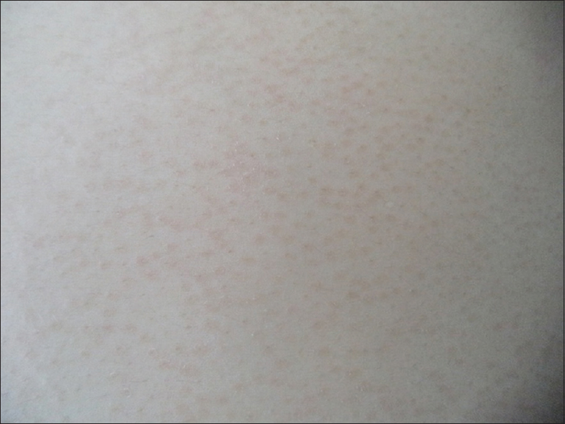
Figure 2: Close view of follicular macules of pityriasis versicolor
A written consent for the examination and biospy was obtained from the patient after having been informed about the procedure.
She was treated with topical sertaconazole 2% cream twice daily and oral fluconazole 400mg weekly for two weeks. After four weeks she was free of skin lesions.
DISCUSSION
Currently Malassezia globosa and Malassezia furfur are the predominant species implicated in pityriasis versicolor [5]. Various species of genus Malassezia are among the normal flora of yeast on human skin. In most cases it is the shift in the relationship between the resident yeast flora and the human skin which presents clinically as pityriasis versicolor. Clinically patients present with well defined discrete or confluent macules characterized by fine branny scaling which can be made prominent by stretching the skin [1]. The various clinical variants of pityriasis versicolor that have been described include hypochromic, hyperchromic, combination of hypochromic and hyperchromic, erythematous, circinate, acral, atrophic, etc. but follicular type has rarely been reported [2–4]. Framil VMS et al in a study of 102 patients of pityriasis versicolor from Brazil reported only one case of follicular variety [2]. The present case was reported due to the rarity of this clinical variant. The various treatment options for pityriasis versicolor include topical azole antifungals, 2.5% selenium sulphide in a detergent base, terbinafine 1% cream, 20% sodium hyposulphite solution, etc. The systemic antifungals which are effective in pityriasis versicolor include fluconazole, ketoconazole, itraconazole, etc. [1]. Our case also responded well to the topical azole (sertaconazole) together with oral fluconazole.
CONSENT
The examination of the patient was conducted according to the Declaration of Helsinki principles. Written informed consent was obtained from the patient for publication of this article and any accompanying images.
REFERENCES
1. Hay EJ, Ashbee HR, Burns T, Breathnach S, Cox N, Griffiths C, MycologyRook’s Textbook of Dermatology 2010; 2: 8th edition. U.K: Wiley-Blackwell; 36.10-36.12.
2. Framil VM, Melhem MS, Szeszs MW, Corneta EC, Zaitz C, Pityriasis versicolor: isolation and identification of the main speciesAn Bras Dermatol 2010; 85: 111-4.
3. Akaberi AA, Amini SS, Hajihosseini H, An Unusual Form of Tinea Versicolor: A Case ReportIran J Dermatol 2009; 12: SupplS30-1.
4. Yang YS, Shin MK, Haw CR, Atrophying Pityriasis Versicolor: Is This a New Variant of Pityriasis Versicolor?Ann Dermatol 2010; 22: 456-9.
5. Gaitanis G, Velegraki A, Alexopoulos EC, Chasapi V, Tsigonia A, Katsambas A, Distribution of Malassezia species in pityriasis versicolor and seborrhoeic dermatitis in Greece. Typing of the major pityriasis versicolor isolate M. globosaBr J Dermatol 2006; 154: 854-9.
Notes
Source of Support: Nil
Conflict of Interest: None declared.
Comments are closed.