Our Dermatol Online. 2012; 3(4): 304-307
Date of submission: 13.07.2012 / acceptance: 21.08.2012
Conflicts of interest: None
A STUDY ON NON VENEREAL GENITAL DERMATOSES IN NORTH INDIA
Neerja Puri, Asha Puri
Department of Dermatology and Venereology, Punjab Health Systems Corporation, Ferozepur, Punjab, India
Corresponding author: Dr. Neerja Puri e-mail: neerjaashu@rediffmail.com
How to cite an article: Puri N, Puri A. A study on non venereal genital dermatoses in north India. Our Dermatol Online. 2013; 4(1): 304-307.
Abstract
Non-venereal dermatoses of female external genitalia include a spectrum of diseases with varied etiology. The most common non-venereal dermatoses in males were scrotal dermatitis seen in 16.6% patients, vitiligo was seen in 14.3 % patients, fixed drug eruption ,scabies and pearly penile papules were seen in 10% patients each. Sebaceous cyst, tinea, psoriasis and lichen planus was seen in 6.6% patients each. Balanitis xerosus obliterans, squamous cell carcinoma and verrucous carcinoma and achrochordon were seen in 3.3% patients each. The most common genital dermatoses seen in females were lichen sclerosus (15%), vitiligo (15%) and vulval candidiasis in 15% cases. Other non venereal genital dermatoses in females were lichen simplex atrophicus (10%), bartholin cyst (10%), tinea (10%), psoriasis (10%)\, vulval lymphoedema (10%) and achrochordon in 5% patients.
Key words: genital; non venereal; female; male; dermatoses
Introduction
Dermatoses involving female external genitalia are not always sexually transmitted. Those which are not sexually transmitted are referred to as non- venereal dermatoses. The various non-venereal dermatoses of female includes inflammatory cutaneous disorders (psoriasis, seborrheic dermatitis, lichen planus, lichen sclerosus), autoimmune (vitiligo), multisystem diseases (Behcet syndrome, Reiter syndrome, Crohn disease), exogenous (contact dermatitis, corticosteroid abuse, fixed drug eruption), and benign and malignant neoplasms (extramammary Paget disease) [1-3]. The non venereal dermatoses in males encompasses two group of disorders [4,5]. Group one consists of disorders that are seen only in the genitalia e.g. angiokeratoma of Fordyce, median raphe cyst), group two comprises of disorders that affect genitalia as well as other parts of the body. Because venereal and non-venereal dermatoses tend to be confused, the occurrence of these dermatoses may be associated with mental distress and guilt feelings in affected patients.
The non venereal dermatoses may be classified into five types based on pathogenesis. It includes inflammatory diseases, infections and infestations, congenital disorders, benign abnormalities, premalignant and malignant lesions. Since these groups include a wide variety of disorders, the identification and establishment of the nature of disease is a challenging venture.it is also important to distinguish between venereal and non venereal dermatoses, as venereal diseases are of primary concern to the patient.
Material and Methods
We selected 50 cases of non venereal genital dermatoses for the study from the department of dermatology and STD. Informed consent was taken from all the patients for the study and prior approval of the hospital ethical committee was taken for the study. Patients having venereal disease were excluded from the study. A detailed history including demographic data, chief complaints related to skin, presence of itching, skin lesions, onset, pregnancy status, menstrual status, and associated medical or skin disorders was elicited and recorded. Enquiry was made with regard to history of sexual exposure. Cases having venereal diseases were excluded from the study.The external genitalia were examined and findings were noted. A detailed physical examination was made to see any associated lesions elsewhere in the body. Investigations such as Gram stain and KOH mount were done as and when required to establish the diagnosis. Biopsy and histopathological examination of the specimen was done when required to confirm the diagnosis. VDRL and Elisa test for HIV were done in all the patients to exclude any sexually transmitted disease.
Results (Tabl I, II)
A total of 50 patients with non venereal genital dermatoses were included in the study. The data was collected and the results were analyzed. The mean age of the patients in our study was 38 years and the commonest age group of the patients was between 30-40 years of age. The majority of the patients were married (96%) and 4% were unmarried. Males outnumbered females in the ratio of 1.5:1.
| Sr no |
Genital dermatoses |
Number |
Percentage |
| 1 |
Vitiligo |
4 |
14.3 |
| 2 |
BXO |
1 |
3.3 |
| 3 |
Scrotal Dermatitis |
5 |
16.6 |
| 4 |
Sebaceous cyst |
2 |
6.6 |
| 5 |
Fixed drug eruption |
3 |
10 |
| 6 |
Tinea |
2 |
6.6 |
| 7 |
Pearly penile papules |
3 |
10 |
| 8 |
Psoriasis |
2 |
6.6 |
| 9 |
Lichen planus |
2 |
6.6 |
| 10 |
Scabies |
3 |
10 |
| 11 |
SCC |
1 |
3.3 |
| 12 |
Verrucous carcinoma |
1 |
3.3 |
| 13 |
Achrochordon |
1 |
3.3 |
| 14 |
TOTAL |
30 |
100 |
Table I. Genital dermatoses in males (n=30)
| Sr no |
Genital dermatoses |
Number |
Percentage |
| 1 |
LSA |
3 |
15 |
| 2 |
LSC |
2 |
10 |
| 3 |
Vitiligo |
3 |
15 |
| 4 |
Vulval candidiasis |
3 |
15 |
| 5 |
Bartholin cyst |
2 |
10 |
| 6 |
Tinea |
2 |
10 |
| 7 |
Psoriasis |
2 |
10 |
| 8 |
Vulval lymphoedema |
2 |
10 |
| 9 |
Achrochordon |
1 |
5 |
| 14 |
TOTAL |
20 |
100 |
Table II. Genital dermatoses in females
Discussion
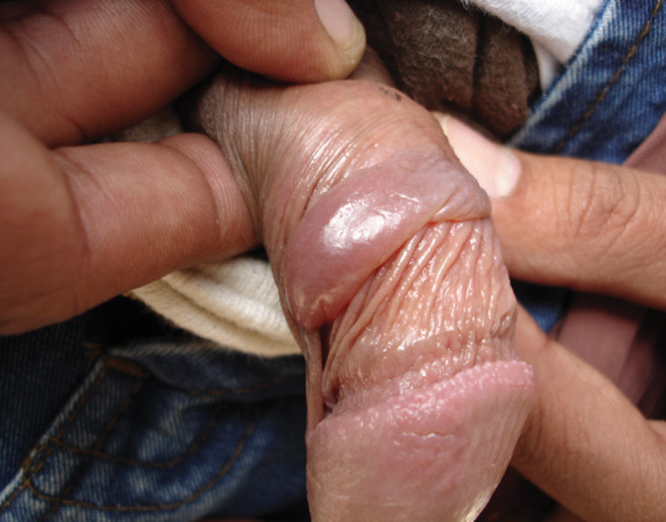
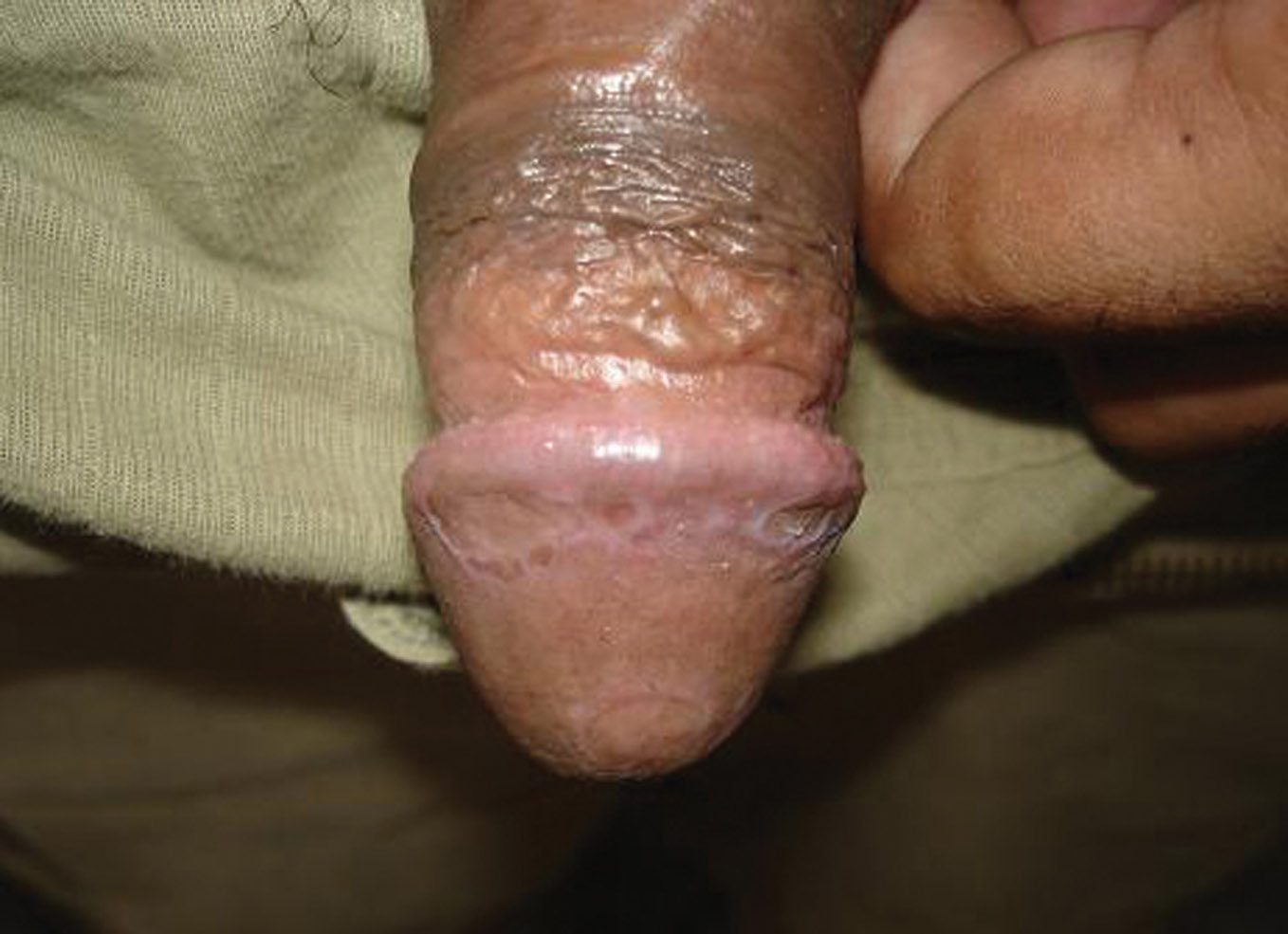
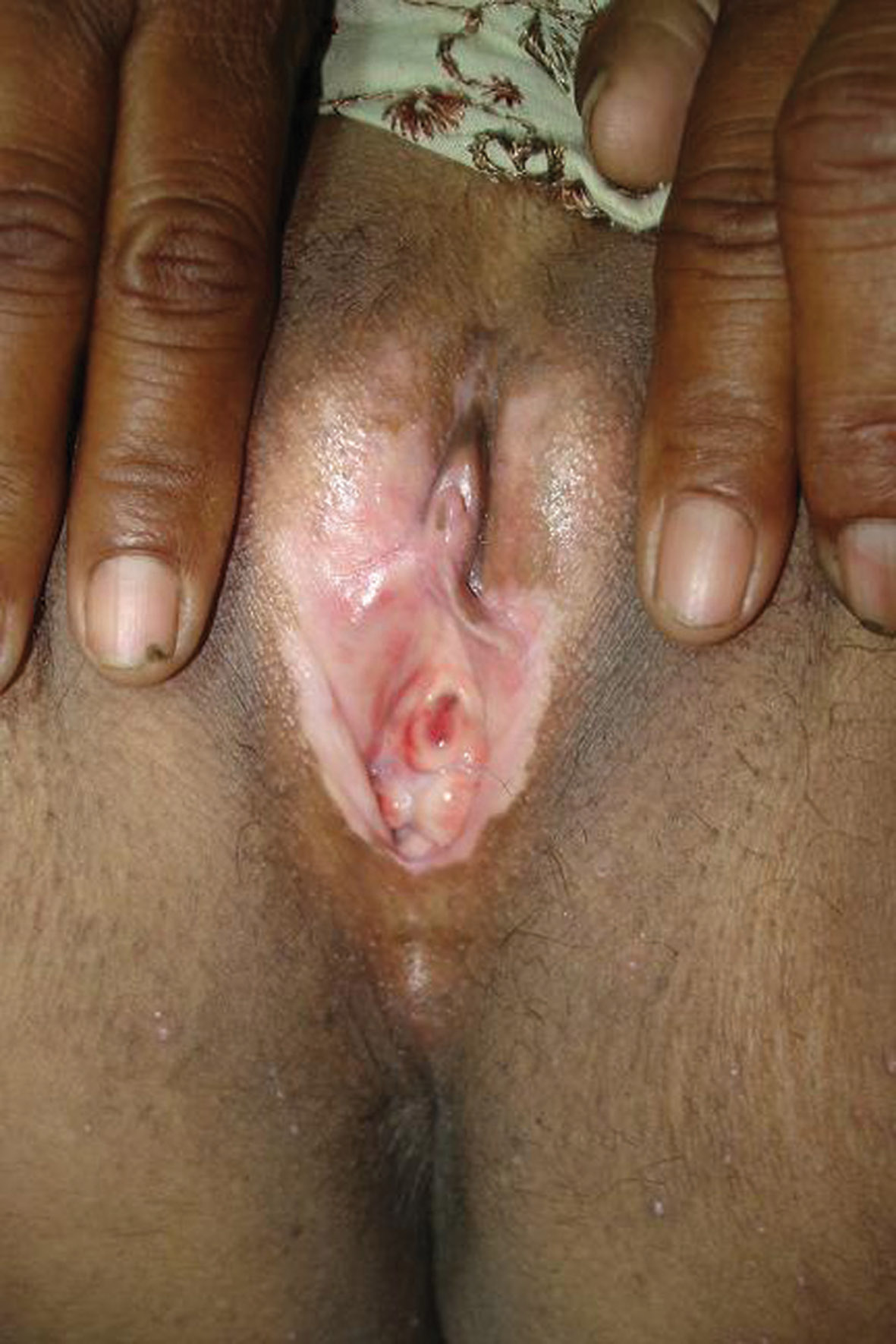
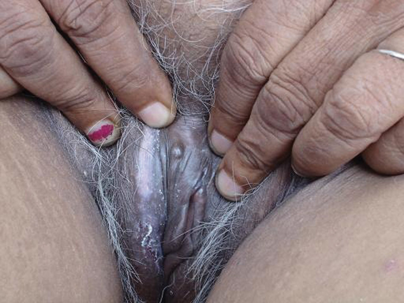
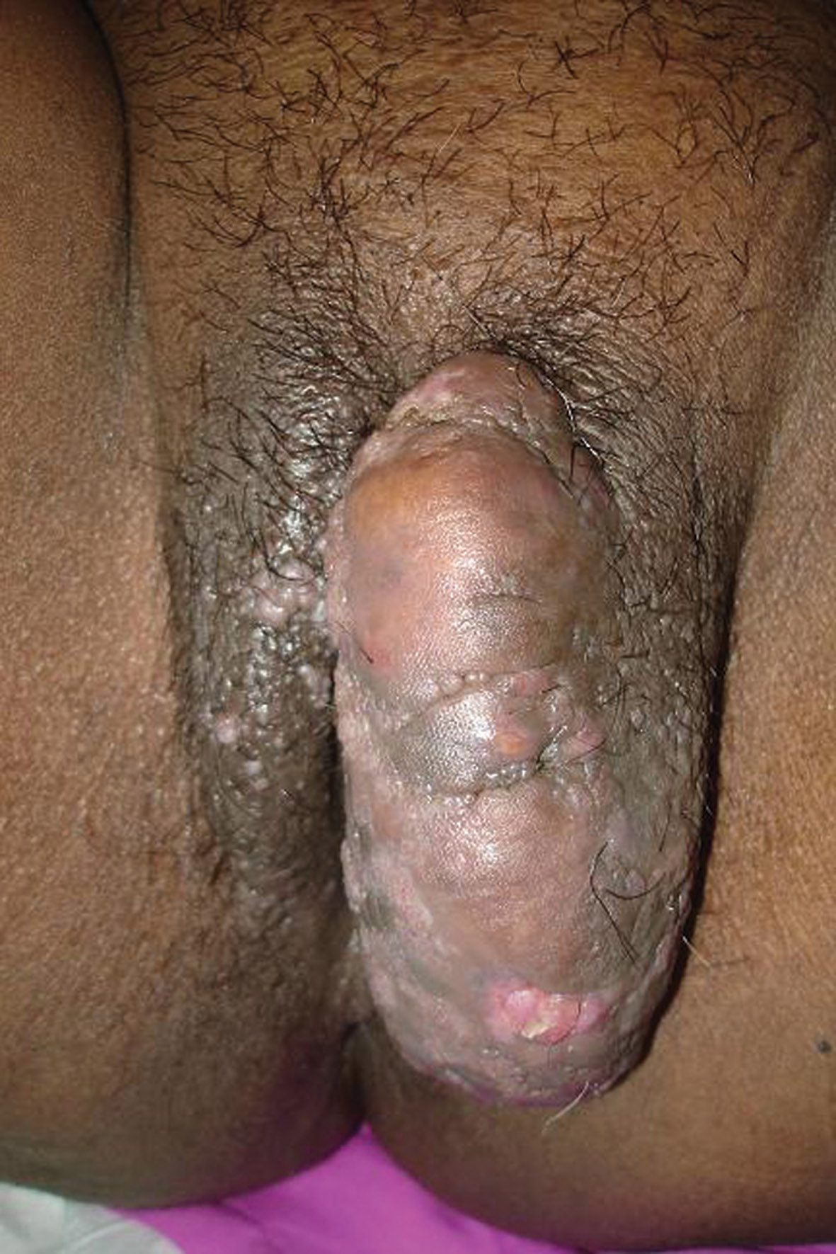
Genital diseases may be associated with severe psychological trauma and fear in the mind of patients. Majority of our patients belonged to age group of 21-40 years.The mean age of the patient was 32 years. Males (60%) outnumbered females (40%) and male : female was 1.5:1. The common presenting feature were itchy genitalia, white discoloration, swelling, pain, burning sensation, mass, dyspareunia, redness, exfoliation of skin, raised lesions over skin, oozing, constipation, burning micturition, ulceration, erosion and thickening of skin. Some patients had more than one complaint. In females, labia majora was the most common site of involvement, accounting for 87% cases followed by labia minora in 48% cases and mons pubis in 10% cases. In males, penis (52%) was the common site of involvement, followed by prepuce (32%) and scrotum in 20 % cases. The most common non-venereal dermatoses in males were scrotal dermatitis seen in 16.6% patients, vitiligo was seen in 14.3 % patients, fixed drug eruption (Fig. 1), scabies and pearly penile papules were seen in 10% patients each. Sebaceous cyst, tinea, psoriasis and lichen planus (Fig. 2) was seen in 6.6% patients each. Balanitis xerosus obliterans, squamous cell carcinoma (Fig. 3) and verrucous carcinoma and achrochordon were seen in 3.3% patients each. The most common genital dermatoses seen in females were lichen sclerosus (Fig. 4) seen in 15% cases, vitiligo (15%) and vulval candidiasis in 15% cases. Other non venereal genital dermatoses in females were lichen simplex atrophicus (Fig. 5) in 10% patients, bartholin cyst (10%), tinea (10%), psoriasis (10%), vulval lymphoedema (Fig. 6) in 10% patients and achrochordon in 5% patients.
|
Figure 1. Bullous Fixed drug eruption in a 25 year old male
|
Figure 2. Lichen planus in a 27 years old male
|
|
Figure 3. Squamous cell carcinoma in a 46 year old male
|
Figure 4. LSA in a 45 year old female
|
|
Figure 5. Lichen simplex atrophicus in a 48 years old female
|
Figure 6. Vulval lymphoedema in a 35 years old female
|
Lichen sclerosus in females is chronic inflammatory dermatoses associated with substantial discomfort and morbidity [7,8]. Anogenital LS is characterized by porcelain white atrophic plaques that may become confluent extending around vulval and perianal skin in a figure-of-eight configuration. The resulting atrophic plaque may have a cellophane-paper-like texture, wrinkled and fragile surface associated with telangiectasia, purpura, erosions, fissuring or ulceration [9,10]. Vitiligo is an acquired pigmentary disorder characterized by loss of melanocytes resulting in depigmentation [11]. Approximately 0.1 percent to 4 percent of people worldwide are affected by vitiligo. Indian studies report 0.46 percent to 8.8 percent prevalence of vitiligo. Lymphedema is swelling attributed to accumulation of lymph in tissue. It is associated with inadequate lymphatic drainage. If it is from intrinsic abnormality of lymph conducting pathways, then it is called primary lymphedema. Primary lymphedema usually involves the lower extremities. But if inadequate lymphatic drainage is from an acquired obstruction or obliteration of lymphatic channels, it is said to be secondary lymphedema. The common causes of secondary lymphedema are trauma (surgery, radiotherapy), infection (filariasis, tuberculosis), inflammation, and malignancy [12]. Vulval lichen planus usually presents as violaceous or erythematous papules or annular plaques or erosions with or without a lacy white border [13,14]. These lesions may ulcerate. If only vulval involvement is present, then disease is more likely to be erosive, with most lesions around labia minora, clitoris and clitoral hood [15]. Bartholin cysts are the most common cystic growths of the vulva [16]. Two percent women develop Bartholin duct cyst or gland abscess at sometime in life. Bartholin gland abscesses are almost three times more common than Bartholin duct cysts [17]. Gradual involution of the Bartholin glands occurs by 30 years of age. This is probably responsible for the more frequent occurrence of Bartholin duct cysts and gland abscesses during the reproductive years, especially between 20 and 29 years of age. It may start as an asymptomatic unilateral nontender cystic swelling, but it can cause pain and limitation of activity with increase in size.
REFERENCES
1. Khaitan BK: Non-venreal diseases of genitalia. In: Sharma VK, editor. Sexually Transmitted Diseases and AIDS. 1st edn. New Delhi: Viva books Pvt Ltd. 2003:413-21.
2. Lynch PJ, Moyal-Barrocco M, Bogliatto F, Micheletti L, Scurry J: Classification of vulvar dermatoses: pathologic subsets and their clinical correlates. J Reprod Med. 2007;52:3-9.
3. Sullivan AK, Straghair GJ, Marwood RP, Staughton RC, Barton SE: A multidisciplinary vulva clinic: the role of genitor-urinary medicine. J Eur Acad Dermatol Venereol. 1999; 13:36-40.
4. Bunker CB, Neill SM: The genital, perianal and umbilical regions. In: Burns T, Breathnach S, Cox N, Griffiths C, editors. Rook’s textbook of dermatology. 7th edn. Oxford: Blackwell Science; 2004:68.1-68.104.
5. Mortimer PS: Disorders of lymphatic vessels. In: Burns T, Breathnach S, Cox N, Griffiths C, editors. Rook’s Textbook of Dermatology, 7th edn. Oxford: Blackwell Science; 2004:51.1- 51.27.
6. Sehgal VN, GAngarani OP: Genital fixed drug eruption. Genitourin Med. 1986;62:56-8.
7. Funaro D: Lichen sclerosus: a review and practical approach. Dermatol Ther. 2004;17:28-37.
8. Powell JJ, Wojnarowska F: Lichen sclerosus. Lancet. 1999;353:1777-83.
9. Powell JJ, Wojnarowska F, Hollowood K: What is the incidence of lichen sclerosus? Br J Dermatol. 2000;143:30.
10. Tasker GL, Wojnarowska F: Lichen sclerosus. Clin Exp Dermatol. 2003;28:128-33.
11. Hillard PJA: Benign diseases of the female reproductive tract: symptoms and signs. In: Berek JS, editor. Novak’s Gynecology.13th edn. Philadelphia: Lippincott Williams and Wilkins; 2002:351-420.
12. Acharya KM, RAnapara H, Sakia JJ, Kaur C: Study of 200 cases of genital lesions of non venereal origin. Ind J Dermatol Venereol Leprol. 1999;64:68-70.
13. Soutter WP: Benign disease of vulva and vagina. In: Shaw RW, Soutter WP, Stanton SL. Editors, Gynaecology. 2nd edn. Edinburgh: Churchill livingstone; 1997:557-68.
14. Pincus SH: Non-venereal disease of the female genitalia. In: Moschella SL, Hurley HJ, editors. Dermatology. 3rd edn. Philadelphia: Saunders Company. 1992:1016-28.
15. Dogra S: Late onset vitiligo: A study of 182 patients. Int J Dermatol. 2005;44:193-6.
16. Omole F, Simmons BJ, Hacker Y: Management of Bartholin duct cyst and gland abscess. Am Fam Physician. 2003;68:135-40.
17. Kaufman RH: Cystic tumors. In: Kaufman RH, Faro S, Brown D, editors. Benign Diseases of the Vulva and Vagina. 5th edition. Philadelphia: Elsevier Mobsy; 2005:216-8.

Comments are closed.